
Why classification names mechanism at the center and organizes probability at the border
Note: The video and audio linked above were generated with the assistance of AI. Clinical accuracy has been reviewed, but no AI-generated content can be guaranteed to be fully error-free.
Why this spoke matters
VWD classification often works remarkably well.
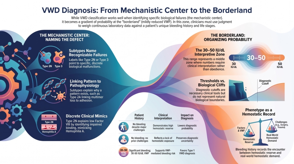
At the mechanistic center of the disease, subtype labels can name recognizable failures in VWF biology. Type 2A points to loss of high-molecular-weight multimers. Type 2B links gain-of-function platelet GPIbα binding to loss of high-molecular-weight multimers and, in some patients, thrombocytopenia. Type 2M points to impaired adhesive function despite relatively preserved multimers. Type 2N points to impaired factor VIII binding. Type 3 points to near-complete absence of VWF.1
That is the strength of classification.
But not every patient stands at the mechanistic center.
Some patients stand near the border, where VWF levels are mildly reduced, bleeding history is variable, family history may be unclear, and laboratory thresholds must do work they were never biologically designed to do.
This essay is about that border.
It is about low VWF, mild type 1 VWD, normal biologic variation, and the clinical judgment required when a continuous trait is divided into diagnostic categories.
The central principle is simple:
At the center, classification names mechanism. At the border, classification organizes probability.
Even that center-to-border distinction is not a cliff. It is a gradient of diagnostic confidence. Some cases name a mechanism clearly. Others require clinicians to weigh probability, context, and risk.
Where classification works well
Classification is easiest when the label points to a distinct biological failure.
If VWF is nearly absent, type 3 VWD is a meaningful category. If VWF cannot bind factor VIII, type 2N explains the low factor VIII and the hemophilia A mimic. If VWF binds platelet GPIbα too avidly, type 2B explains why a gain-of-function interaction can produce bleeding through multimer loss and sometimes thrombocytopenia. If high-molecular-weight multimers are lost, type 2A links structure to platelet adhesion under shear.
In these settings, the subtype does not merely describe a laboratory pattern.
It explains why the pattern exists.
That does not mean every case is simple. Assays can be limited. Phenotypes may overlap. Genotype and phenotype may not align perfectly. Acquired von Willebrand syndrome can mimic inherited disease. But the classification is still doing something powerful: it is linking a name to a mechanism.
The problem becomes harder when the abnormality is not qualitative and discrete, but quantitative and mild.
That is the borderland.
The borderland of mild quantitative VWF deficiency
VWF levels vary continuously across the population.
They are influenced by genetic factors, ABO blood group, age, inflammation, hormonal state, pregnancy, stress, exercise, comorbid illness, and other modifiers.2
That means there is no natural cliff between normal VWF, low VWF, and mild type 1 VWD.
There are values that are clearly abnormal.
There are values that are clearly normal.
And there is a broad middle zone where the number must be interpreted rather than simply obeyed.
This is especially true in the range often described historically as 30 to 50 IU/dL. Some patients in this range have substantial mucocutaneous bleeding. Some do not. Some have family histories that support inherited bleeding risk. Some have no bleeding history because they have never faced a meaningful hemostatic challenge. Some have VWF levels that rise over time. Some have bleeding that persists even after levels normalize.3
The borderland is difficult because the biology is continuous, but clinical care requires decisions.
Thresholds are necessary, but they are not nature
Diagnostic thresholds are not mistakes.
They are necessary.
Clinicians need thresholds to standardize diagnosis, guide testing, communicate risk, design studies, determine eligibility for care, and decide when treatment planning is appropriate.
But thresholds should not be mistaken for natural biological boundaries.
A VWF level of 49 IU/dL is not biologically transformed by becoming 51 IU/dL. A patient at 32 IU/dL does not necessarily have a different kind of biology from a patient at 28 IU/dL. The number matters, but the number does not carry the whole diagnosis.
The 2021 ASH/ISTH/NHF/WFH diagnostic guideline recommends diagnosing type 1 VWD in patients with VWF levels below 0.30 IU/mL regardless of bleeding, and in patients with abnormal bleeding and VWF levels below 0.50 IU/mL.4
That recommendation has practical strengths. The rationale includes improving recognition, reducing diagnostic delay, and helping bleeding patients access care, especially patients whose symptoms have historically been minimized.
It also raises concerns. Some experts worry that expanding type 1 VWD terminology into the 30 to 50 IU/dL range may medicalize people whose mildly reduced VWF level is better understood as a bleeding risk factor than as a monogenic bleeding disorder.5
Both concerns are real.
Thresholds help structure the conversation.
They do not end it.
Low VWF and type 1 VWD
The term low VWF has been used to describe patients with mildly reduced VWF levels, often in the 30 to 50 IU/dL range, especially when the biology does not clearly resemble classic inherited type 1 VWD.
The concept arose because patients in this range are heterogeneous.
Compared with patients with lower VWF levels, they are less likely to have identifiable pathogenic VWF variants, less likely to show clear VWF-linked inheritance, and more likely to involve heterogeneous contributions from VWF, ABO blood group, and other genetic or hemostatic modifiers. Yet referred patients in this range may still have clinically important bleeding.6
This is why low VWF has been described in different ways:
- as a mild form of type 1 VWD
- as a bleeding risk factor
- as a subgroup within heterogeneous type 1 VWD
- as a management category
- as a marker that requires interpretation in clinical context
The disagreement is not merely semantic.
The name affects how patients understand themselves, how clinicians plan procedures, how families are counseled, how research cohorts are built, and how care is accessed.
But the practical question is often not, “Which term wins?”
The practical question is:
How likely is this patient to have clinically meaningful VWF-mediated bleeding, and what should we do before the next hemostatic challenge?
Bleeding phenotype is not noise
At the border, bleeding history becomes central.
A mildly reduced VWF level without bleeding is not the same clinical problem as the same VWF level in a patient with heavy menstrual bleeding since menarche, postpartum hemorrhage, iron deficiency, prolonged bleeding after dental extraction, and a family history of mucocutaneous bleeding.
The number may be similar.
The probability is not.
Bleeding assessment tools can help standardize the history and reduce reliance on vague impressions. They are especially useful when the question is whether bleeding is greater than expected for age and exposure history.7
But bleeding scores are not perfect.
They depend on age, sex, recall, treatment history, and prior opportunity to bleed. A person who has never had surgery, childbirth, dental extraction, or major mucosal injury may have a low score because they are hemostatically normal, or because they have not yet been tested by life.
A bleeding phenotype is not just a symptom list.
It is a record of encounters between hemostatic reserve and hemostatic demand.
The problem of untested hemostatic challenge
A negative bleeding history is meaningful only if the patient has had the opportunity to bleed.
This is one of the most important lessons in the borderland.
A child with mild epistaxis and low-normal VWF may not yet have had dental extraction, surgery, menarche, pregnancy, or trauma. A young man may have no heavy menstrual bleeding or childbirth history by definition and may have avoided major procedures. A patient may appear asymptomatic because life has not yet asked the hemostatic system a difficult question.
In such patients, classification must remain provisional.
That does not mean every person with a borderline VWF level should be labeled as diseased. But it does mean that clinicians should distinguish between:
no bleeding despite meaningful challenges
and
no bleeding because meaningful challenges have not occurred
Those are not the same history.
The first lowers probability.
The second preserves uncertainty.
Age-related normalization
VWF levels often rise with age.
This creates another borderland problem.
A patient diagnosed earlier in life with low VWF or type 1 VWD may later have VWF levels in the normal range. The question then becomes: has the diagnosis disappeared, or has the laboratory value changed?
The answer depends on the whole pattern.
If the original diagnosis was based only on a borderline laboratory value in a patient without bleeding, later normalization may support reconsidering the label. If the patient has had no bleeding despite meaningful challenges, the argument for removing or softening the diagnosis becomes stronger.
But if the patient has a strong lifelong mucocutaneous bleeding history, postpartum hemorrhage, procedure-related bleeding, iron deficiency, and repeatedly low historical VWF values, a normal VWF value later in life should not automatically erase the clinical story.8
The current level matters for current management.
The historical phenotype matters for interpretation.
Both can be true.
Dangers at both edges
The borderland has two dangers.
The first is overdiagnosis.
Overdiagnosis can turn normal biological variation into disease. It can medicalize people unnecessarily, create anxiety, complicate insurance or life planning, expose patients to unneeded treatment, and distort family interpretation.
The second is underdiagnosis.
Underdiagnosis can dismiss real bleeding, delay recognition, leave heavy menstrual bleeding untreated, miss iron deficiency, fail to plan for surgery or childbirth, and deny patients access to hemostatic support.
Both errors matter.
A clinician who is afraid of overdiagnosis may minimize bleeding.
A clinician who is afraid of underdiagnosis may overlabel borderline laboratory values.
Good classification avoids both reflexes.
It asks how well the whole pattern fits.
Evidence anchor: why the borderland is difficult
Summary derived from low VWF cohorts, diagnostic guidelines, expert reviews, and studies of age-related VWF change.
| Evidence stream | What it shows | Why it matters |
|---|---|---|
| Population distribution | VWF levels vary continuously, and mildly reduced values occur in otherwise healthy people | there is no natural biologic cliff at a diagnostic threshold |
| Genetic data | pathogenic VWF variants are detected more frequently at lower VWF levels and less frequently in the 30 to 50 IU/dL range | mild quantitative deficiency is often genetically heterogeneous |
| Bleeding cohorts | referred patients with low VWF can have significant mucocutaneous bleeding | mild laboratory abnormalities can still matter clinically |
| Bleeding correlation | within the 30 to 50 IU/dL range, bleeding severity often correlates poorly with the absolute VWF level | the clinical phenotype cannot be read from the number alone |
| Age-related change | VWF levels often rise with age, sometimes into the normal range | current VWF levels must be interpreted alongside historical bleeding and prior values |
| Guideline evolution | older frameworks often preserved “low VWF” as a separate category, whereas the 2021 guideline classifies bleeding patients below 0.50 IU/mL as type 1 VWD | classification reflects both biology and clinical consequences |
Interpretive note: The borderland is difficult because VWF is a continuous biological trait, bleeding is context-dependent, and diagnostic labels influence care. The question is not only whether the VWF level is low. The question is whether the patient’s overall pattern supports clinically meaningful VWF-mediated bleeding.9
Clinical synthesis
At the mechanistic center of VWD, classification often names the defect.
At the border, classification estimates probability.
Each additional piece of evidence shifts that probability rather than establishing certainty by itself.
That probability is shaped by:
- VWF level
- repeat testing
- bleeding history
- family history
- prior hemostatic challenges
- age
- ABO blood group
- physiologic context
- laboratory pattern
- exclusion of alternative causes
ABO blood group helps explain variation in VWF level, but it should not be used by itself to dismiss clinically meaningful bleeding. ABO-specific diagnostic reference ranges are not recommended in the 2021 ASH/ISTH/NHF/WFH diagnostic guideline.10
The diagnosis is strongest when these elements point in the same direction.
The diagnosis is weakest when it rests on a single borderline value without bleeding, without repeat testing, and without a meaningful hemostatic challenge history.
The borderland does not mean classification has failed.
It means classification has reached the place where judgment is required.
Reflect & Apply
A 42-year-old woman is referred for heavy menstrual bleeding and iron deficiency.
She reports:
- heavy menstrual bleeding since menarche
- postpartum hemorrhage after her first delivery
- prolonged bleeding after wisdom tooth extraction
- easy bruising since adolescence
- no major surgery other than childbirth-related procedures
Her testing, repeated twice when well, shows:
- VWF antigen: 42 IU/dL and 46 IU/dL
- platelet-dependent VWF activity: 39 IU/dL and 44 IU/dL
- factor VIII: mildly reduced
- activity-to-antigen ratio: preserved
- multimers: normal
- platelet count: normal
- no pathogenic VWF variant identified on available testing
Under the 2021 ASH/ISTH/NHF/WFH diagnostic guideline, her abnormal bleeding history and repeatedly reduced VWF levels below 0.50 IU/mL support a diagnosis of type 1 VWD.11
But the biology remains worth thinking through.
Older or alternative frameworks might describe this as low VWF. The preserved activity-to-antigen ratio and normal multimers support a partial quantitative pattern rather than a type 2 qualitative defect. The absence of an identified pathogenic VWF variant does not exclude clinically meaningful bleeding, especially in the 30 to 50 IU/dL range.
What additional history would matter?
What would you want to know before surgery?
Would your answer change if she had never undergone childbirth, dental extraction, or surgery?
Would your answer change if she had no bleeding symptoms despite multiple hemostatic challenges?
The point is not that one label solves the case.
The point is that the label should help organize the probability of future bleeding and the need for planning.
At the border, classification is not a verdict.
It is a structured way to think.
Test your thinking
An interactive quiz for this topic is being developed and will be added soon.