
In this video lecture, Dr. Mackenzie Bowman discusses:
- The advantages and limitations of cellular models used to study von Willebrand factor synthesis, storage, secretion, and function.
- How HUVECs, heterologous expression systems, and patient-derived endothelial colony-forming cells have advanced understanding of VWD pathobiology.
- Emerging cellular approaches for modeling VWD and testing targeted therapies, including iPSC-derived endothelial cells and gene-editing platforms.
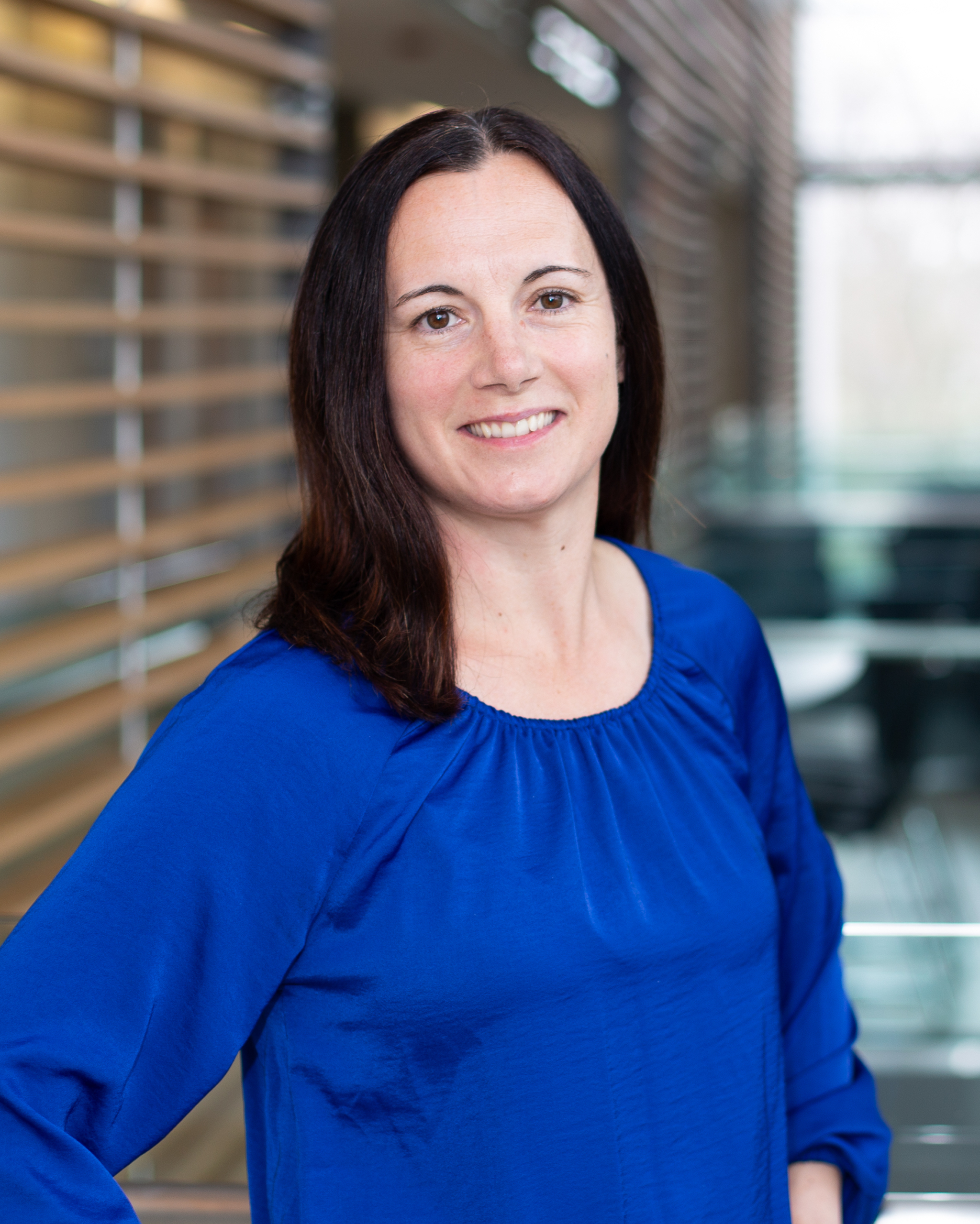
Mackenzie Bowman (PhD) received her PhD from Queen’s University, Kingston, Canada in 2013 with her thesis titled “Investigating the Genetic Basis of Type 3 of von Willebrand Disease (VWD)”. She completed a post-doctoral fellowship in the lab of Dr. Paula James and has continued to work in the James Lab as a Research Associate. Dr. Bowman is also an Assistant Professor (Adjunct) in the Department of Medicine at Queen’s University and is a Senior Clinical Scientist in the Canadian National Inherited Bleeding Disorder Genotyping Laboratory. Over the past 20 years, Dr. Bowman’s research in VWD has included bleeding assessment tools and assessing the prevalence of VWD in primary care, genetics studies, and the use of cellular models for VWD studies, specifically endothelial colony-forming cells (ECFCs).
(Video Lecture Summary)
Introduction
Dr. Mackenzie Bowman reviews the cellular models used to investigate von Willebrand factor (VWF) and von Willebrand disease (VWD). She compares several experimental systems, including primary endothelial cells, heterologous cell lines, endothelial colony-forming cells, induced pluripotent stem cell-derived endothelial cells, and newer gene-edited models. Each model provides distinct opportunities for studying VWF synthesis, secretion, storage, and disease-associated variants. However, each also has limitations that researchers must consider when selecting a model for a particular research question.
Challenges of Studying VWF-Producing Cells
VWF is naturally produced by endothelial cells and megakaryocytes. In endothelial cells, it is stored in Weibel-Palade bodies. In megakaryocytes, VWF is packaged into the alpha granules of platelets. Although these primary cells provide the most physiologically relevant setting for studying VWF, they can be difficult to obtain and maintain.
Megakaryocytes are especially challenging because they represent only a small fraction of nucleated bone marrow cells. Their collection may require an invasive bone marrow biopsy. They can also be generated from circulating CD34-positive hematopoietic progenitor cells, but this requires specialized expertise and produces cells with a limited lifespan. As a result, megakaryocytes and platelets are not commonly used as practical models for VWF investigation.
Primary endothelial cells can be isolated from different vascular beds, but access depends on donor availability and may require invasive procedures. These cells also have limited replicative capacity, creating a need for more accessible and reproducible experimental systems.
Human Umbilical Vein Endothelial Cells
Human umbilical vein endothelial cells, commonly known as HUVECs, have played a foundational role in VWF research. Umbilical cords are generally discarded after delivery, providing a relatively accessible source of primary endothelial cells. The isolation procedure is straightforward and typically produces cells with the characteristic cobblestone appearance of endothelial cultures.
Early HUVEC studies identified VWF within Weibel-Palade bodies and established these structures as important sites of VWF processing and storage. Subsequent experiments using HUVECs helped clarify the stages of VWF biosynthesis, multimerization, storage, and secretion.
These studies also demonstrated that VWF released following endothelial stimulation contains larger multimers and has greater functional activity than constitutively secreted VWF. HUVECs continue to be widely used because they reproduce many important features of endothelial VWF biology.
However, HUVECs reflect neonatal rather than adult endothelial biology. They also have a finite lifespan and must be used at relatively low passage numbers. Access to HUVECs carrying specific patient-associated VWF variants is especially limited.
Heterologous Cell Expression Systems
Heterologous expression systems offer an alternative to primary endothelial cells. In these models, researchers introduce a plasmid containing the VWF gene into non-endothelial cells that do not naturally produce VWF. The cells then express recombinant VWF, allowing investigators to examine protein synthesis, processing, secretion, multimer formation, and intracellular localization.
Early experiments using COS and Chinese hamster ovary cells demonstrated that recombinant VWF could be processed and secreted as high-molecular-weight multimers. These findings established heterologous systems as useful tools for studying the functional effects of specific VWF variants.
Different cell lines vary in their ability to reproduce endothelial features. Some do not form VWF-containing storage organelles. Others, including HEK293 cells, form structures resembling Weibel-Palade bodies, known as pseudo-Weibel-Palade bodies. For this reason, HEK293 cells have become one of the most frequently used heterologous systems in VWF research.
These models allow investigators to study homozygous and heterozygous disease states by expressing mutant VWF alone or together with wild-type VWF. They are particularly useful for defining how individual variants affect VWF biology.
Nevertheless, heterologous systems lack many features of true endothelial cells. Some commonly used cell lines are non-human, VWF expression is typically temporary, and researchers must first generate plasmids carrying the variants they wish to study. Stable expression requires additional methods such as viral transduction.
Endothelial Colony-Forming Cells
Endothelial colony-forming cells, or ECFCs, provide a more physiologically relevant and personalized model. These cells originate from circulating endothelial progenitors and can be isolated from peripheral blood. This allows researchers to obtain patient-specific endothelial cells through a relatively non-invasive blood draw.
Following isolation and culture, ECFC colonies develop the typical cobblestone morphology of endothelial cells. Their identity can be confirmed through endothelial markers, absence of hematopoietic markers, and functional assays such as capillary-like structure formation.
ECFCs derived from patients naturally carry the VWF variant of interest and often reproduce the corresponding quantitative or qualitative VWF defect. Researchers can examine intracellular VWF, constitutive and stimulated secretion, Weibel-Palade body structure, multimer patterns, and responses to secretagogues. These results can also be compared with findings from the same patient’s plasma.
ECFCs and VWD Pathobiology
ECFC studies have provided important insights into the mechanisms underlying VWD. Patient-derived cells have helped identify pathogenic processes such as retention of abnormal VWF within the endoplasmic reticulum and impaired secretion.
These cells have also contributed to understanding the relationship between VWF and angiogenesis. Studies comparing ECFCs from healthy individuals with those from patients with VWD have identified differences in endothelial proliferation, migration, and vessel-like tube formation. However, Dr. Bowman emphasizes that these phenotypes vary among patients and VWD subtypes.
The growing use of ECFCs led the International Society on Thrombosis and Haemostasis to recommend standardized terminology and culture practices. Multicenter studies have also examined normal variation among ECFCs from healthy donors. These investigations identified substantial differences not only between individuals but also between separate ECFC clones obtained from the same person.
This underlying heterogeneity must be understood before patient-derived cells can be interpreted as reliable disease models.
Testing Targeted Therapies in Patient-Derived Cells
ECFCs are also being used to evaluate targeted therapeutic strategies. Dr. Bowman reviews studies using allele-specific approaches to suppress pathogenic VWF variants while preserving expression from the healthy allele.
In one approach, small interfering RNA was designed to target a single nucleotide polymorphism located on the same allele as a dominant-negative type 2A VWF variant. Selective suppression of that allele reduced endoplasmic reticulum retention and improved the cellular phenotype.
A more recent study applied CRISPR-Cas9 gene editing to selectively disrupt pathogenic alleles in ECFCs derived from patients with type 2A and type 2B VWD. Targeting the disease-associated allele improved the cellular phenotype, while targeting the healthy allele worsened it. This work provided proof of principle for an allele-selective gene-editing strategy that could potentially be applied across multiple VWD subtypes.
Limitations of ECFC Models
Despite their advantages, ECFCs remain primary cells with limited proliferative capacity. Isolation success can be low or inconsistent because the progenitor cells are rare in peripheral blood. Patient age and underlying disease may also affect the likelihood of successful isolation.
Because ECFCs retain the complete genetic background of each donor, differences unrelated to the VWF variant may influence experimental results. Variation between separate clones from the same individual can further complicate interpretation.
Induced Pluripotent Stem Cell Models
Patient-derived induced pluripotent stem cells can be differentiated into endothelial cells and may overcome some limitations of primary models. They can self-renew, preserve patient-specific genetic information, and potentially provide a renewable source of endothelial cells.
However, current iPSC-derived endothelial cells produce substantially less VWF than primary endothelial models such as HUVECs or ECFCs. Their Weibel-Palade bodies also tend to be smaller and more rounded, suggesting an immature endothelial phenotype. Efforts to improve endothelial maturation have produced only partial improvements, and the process remains costly and time-consuming.
Novel Gene-Edited and Organ-Specific Models
Dr. Bowman describes a newer approach in which researchers used CRISPR-Cas9 to remove VWF from cord blood-derived ECFCs. Investigators could then reintroduce wild-type VWF or selected VWF variants into a physiologically relevant endothelial environment.
This system reproduced patient-associated defects in VWF processing and generated authentic Weibel-Palade bodies. It also modeled disease phenotypes more accurately than a HEK293 expression system. The platform may be useful for studying VWF glycosylation, storage organelle formation, stimulated secretion, angiogenesis, and additional aspects of VWF biology.
The lecture also considers the potential value of organ-specific endothelial models. Because heavy menstrual bleeding is common among women with VWD, endometrial endothelial cells may help researchers examine how VWF functions within the uterine environment. Dr. Bowman notes the emerging possibility of isolating relevant cells from menstrual blood rather than through an invasive endometrial biopsy.
Conclusion
Dr. Bowman concludes that cellular models have been essential for understanding VWF synthesis, processing, storage, secretion, and the molecular pathobiology of VWD. HUVECs, heterologous cell systems, ECFCs, iPSC-derived endothelial cells, and newer gene-edited models each offer distinct experimental strengths.
No single model is appropriate for every research question. Careful consideration of physiologic relevance, accessibility, reproducibility, cellular maturity, and genetic background is therefore essential. As these platforms continue to advance, they will support deeper investigation of VWD mechanisms and the development of increasingly targeted therapeutic approaches.