When the DAT Keeps Changing
When the DAT Keeps Changing
Cold, warm, or mixed autoimmune hemolytic anemia in critical illness
By William C. Aird, MD
Case Presentation
A 71-year-old woman was transferred from an outside hospital for evaluation of severe hemolytic anemia.
Her medical history included:
- hypertension
- multiple revisions of a left knee prosthesis
- recent placement of an antibiotic spacer for presumed joint infection
Three days before presentation she developed:
- diffuse body aches
- worsening fatigue
- progressive weakness
Laboratory testing at the outside hospital showed profound anemia, representing a hematologic emergency in which stabilization and transfusion support had to proceed in parallel with diagnostic evaluation.
| Test | Result |
|---|---|
| Hemoglobin | 3.5 g/dL |
| LDH | 2152 U/L |
| Total bilirubin | 3.0 mg/dL |
| WBC | 43 ×10⁹/L |
The automated CBC analyzer was unable to report RBC indices because of marked red cell clumping.
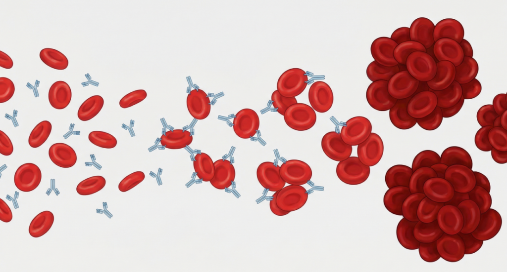
Peripheral smear demonstrated prominent red cell agglutination, raising concern for cold agglutinin disease.
Direct antiglobulin testing performed at the outside hospital suggested:
- cold autoantibody present
- anti-IgG negative
Which laboratory feature most strongly suggests cold agglutinin–mediated hemolysis?
Explanation
Cold agglutinin disease is characterized by IgM antibodies that bind red cells at lower temperatures, causing:
- red cell agglutination
- complement activation
- hemolysis
Agglutination may occur in vivo in cooler peripheral circulation and in vitro when blood samples cool after collection, producing the striking clumping seen on the peripheral smear.
Agglutination may be so prominent that automated analyzers cannot measure RBC indices, producing messages such as “unable to report.”
By contrast, elevated bilirubin and LDH are nonspecific markers of hemolysis, and the leukocytosis in this case raised a separate differential that included severe physiologic stress, infection, and systemic inflammation.
Clinical Course
At this stage, the mechanism of hemolysis was still uncertain. Although the smear suggested cold agglutinin disease, C3 positivity on the DAT can also be seen in warm autoimmune hemolytic anemia, because IgG-mediated antibodies may activate complement, and the IgG can dissociate from red cells during testing.
Definitive distinction requires additional testing, including cold agglutinin titers and thermal amplitude studies, which were pending.
The patient received multiple transfusions and was treated with:
- corticosteroids
- rituximab
- IVIG
Corticosteroids and IVIG are typically ineffective in isolated cold agglutinin disease. However, when the mechanism of autoimmune hemolysis is uncertain at presentation, clinicians may initiate these therapies empirically while additional antibody characterization is underway.
Her hemoglobin initially stabilized.
However, over the following days her clinical course became complicated by:
- septic shock
- acute liver injury
- need for mechanical ventilation
- continuous renal replacement therapy
At the same time, repeat serologic testing became more difficult to interpret because the patient had now received multiple transfusions, corticosteroids, rituximab, and IVIG, all of which could alter the apparent immunohematologic picture.
During hospitalization, repeat DAT testing later showed:
- 1+ anti-IgG
- no anti-C3 activity
What does this new pattern most strongly suggest?
Explanation
Warm autoimmune hemolytic anemia is typically characterized by:
- IgG-mediated antibodies
- DAT positive for IgG
In contrast, cold agglutinin disease typically shows:
- C3 positivity
- little or no IgG
The shift in DAT pattern suggested that the patient may have had:
- mixed autoimmune hemolytic anemia, or
- evolution from cold-predominant to warm-predominant hemolysis
At the same time, this result had to be interpreted cautiously because transfusions and immunomodulatory therapies can introduce substantial noise into DAT interpretation.
Diagnostic Challenges in Critical Illness
Interpreting hemolysis markers became difficult because the patient developed severe liver injury.
Several laboratory findings were therefore difficult to interpret:
| Marker | Interpretation challenge |
|---|---|
| Low haptoglobin | may reflect liver failure rather than hemolysis |
| Hyperbilirubinemia | predominantly direct bilirubin from liver injury |
| Hyperferritinemia | may reflect systemic inflammation or liver failure |
Peripheral smear demonstrated:
- numerous nucleated red cells
- polychromasia
- rouleaux formation
These findings suggested severe marrow stress in the setting of prior massive hemolysis and critical illness.
When standard hemolysis markers become unreliable, clinicians must rely more heavily on:
- serial hemoglobin and hematocrit trends
- smear morphology
- transfusion requirements
- the overall clinical trajectory
Which feature of this case best illustrates the diagnostic complexity of autoimmune hemolytic anemia in critically ill patients?
Explanation
Autoimmune hemolytic anemia can evolve during the course of illness.
Some patients develop mixed AIHA, characterized by:
- warm IgG antibodies
- cold complement-activating antibodies
In critically ill patients, interpretation becomes even harder because of:
- liver dysfunction
- systemic inflammation
- multiple transfusions
- immunosuppressive therapy
- critical illness–related marrow stress
The challenge is not simply to assign a label, but to decide which mechanism is dominant now, and whether treatment should be directed primarily at cold agglutinin biology, warm AIHA, or both.
Management
The patient was treated with:
- prednisone 1 mg/kg/day
- rituximab (initial dose given)
- IVIG
Because of uncertainty regarding the dominant mechanism of hemolysis, rituximab was held temporarily while further blood bank testing was performed.
This additional testing was intended to better characterize the antibody pattern and clarify whether the clinically relevant process remained:
- predominantly cold-mediated
- predominantly warm-mediated
- or mixed
Supportive care included:
- warmed blood transfusions
- frequent hematocrit monitoring
- treatment of underlying critical illness
The decision to hold rituximab reflected an important principle: treatment target matters. B-cell–directed therapy is rational for cold agglutinin–mediated disease, but when the dominant mechanism is uncertain in a severely ill patient, clinicians may need to pause and reassess before proceeding further.
Teaching Points
- Autoimmune hemolytic anemia can present with profound anemia.
Severe hemolysis may produce hemoglobin levels below 4 g/dL and interfere with automated CBC analysis. - Red cell agglutination on smear strongly suggests cold agglutinin–mediated hemolysis.
Agglutination may cause analyzer failure and spurious laboratory results. - Some patients develop mixed warm and cold autoimmune hemolytic anemia.
DAT patterns may change over time as different antibodies become dominant. - Critical illness can obscure the interpretation of hemolysis markers.
Liver failure, inflammation, transfusions, and immunotherapy may complicate assessment of ongoing hemolysis.
Why This Case Matters
Autoimmune hemolytic anemia is often presented as a static diagnostic category.
In practice, the disease can evolve over time.
Patients may move through several stages:
- initial massive hemolysis
- mixed antibody patterns
- changing DAT results
- diagnostic uncertainty during critical illness
This case shows that the hardest part is not simply naming the disease. It is deciding, in real time, which mechanism is driving hemolysis now and whether treatment should be adjusted accordingly.
Recognizing that AIHA can shift in mechanism during the course of illness helps clinicians avoid premature diagnostic closure and adapt management as new information emerges.