How Does Iron Get Into and Out of Ferritin?
Schematic overview
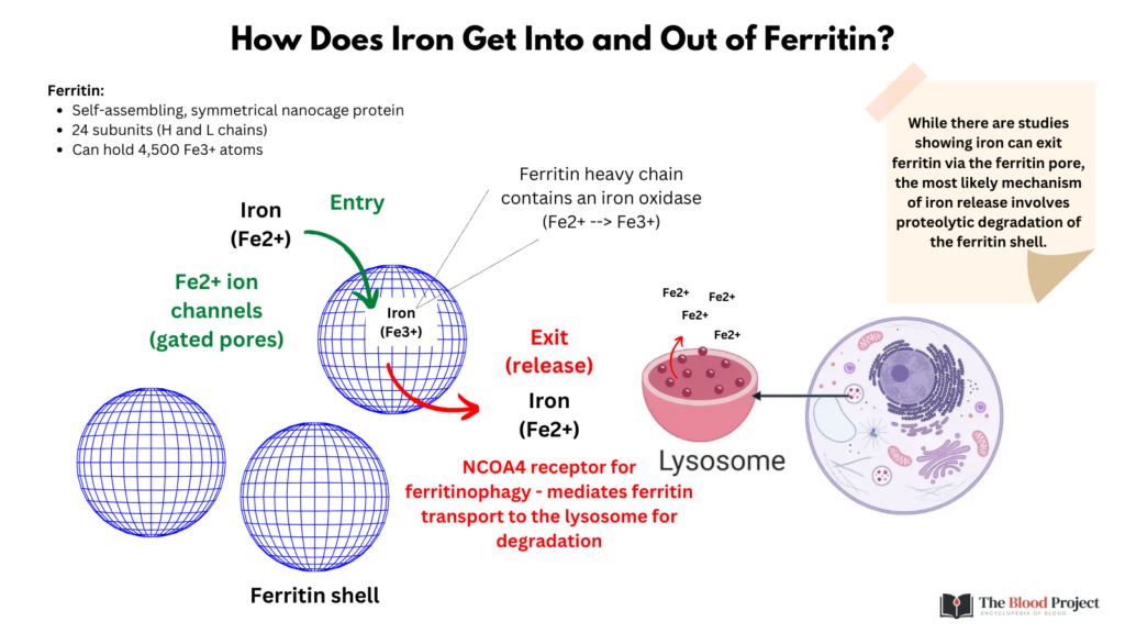
How does iron enter the ferritin shell?
- Ferrous ions (Fe2+) can diffuse into the core and enter the ferritin protein cage through 8 hydrophilic Fe2+ ion channels at the 3-fold symmetry axis, where they are oxidised by dioxygen (or H2O2 if present) at a di-iron catalytic site to form Fe(III)2–O products that then form the Fe2O3H2O.1
How does iron exit the ferritin shell?
- The mechanism underlying the release of iron release from ferritin is called ferritinophagy, which is a form of autophagy.
- Autophagy:2
- A highly conserved process (present in all eukaryotes) that leads to degradation of cytoplasmic organelles, proteins, and macromolecules, and the recycling of the breakdown products.
- In mammalian cells, there are three primary types of autophagy:3
- Microautophagy
- Macroautophagy
- Chaperone-mediated autophagy (CMA)
- All three types of autophagy culminate in the delivery of cargo to the lysosome for degradation and recycling.
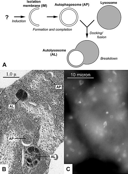
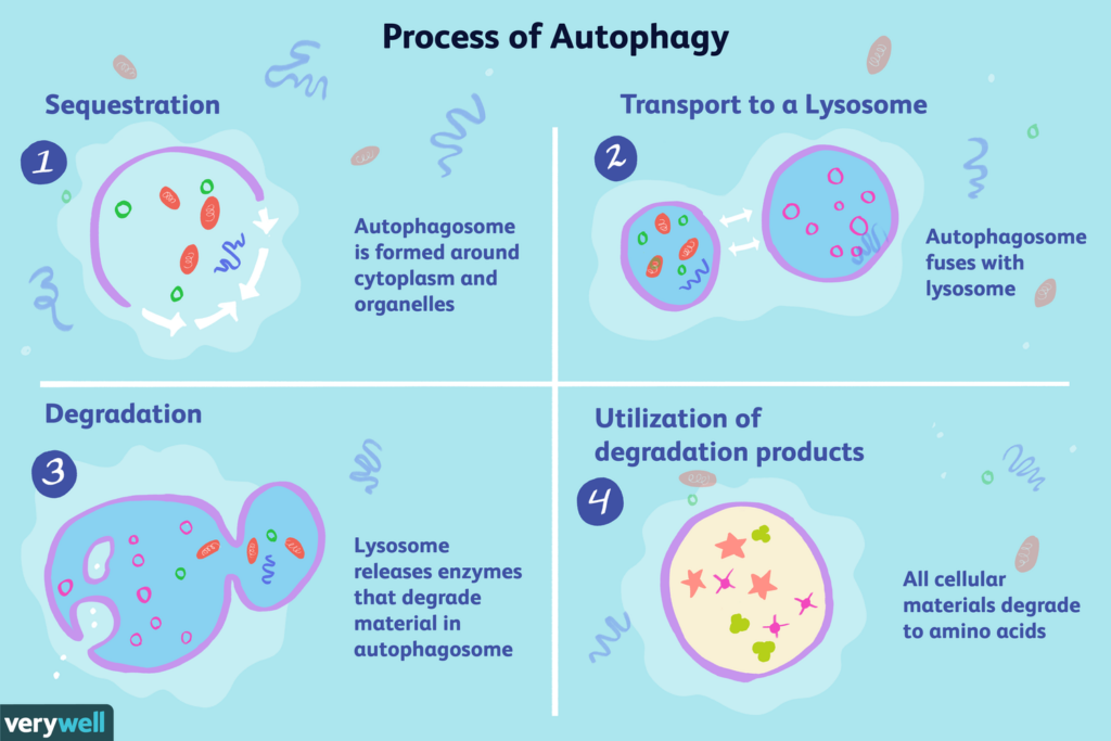
- The process of macroautophagy (of which ferritinophagy is an example) involves four sequential steps:4
- Sequestration: The synthesis of double-membrane sequestering vesicles—autophagosomes—is used to sequester cargo and subsequently transport it to the lysosome.
- Transfer: This takes place when the autophagosome is fused to a lysosome. Lysosomes contain enzymes that degrade dysfunctional components.
- Degradation: This happens when a lysosome releases enzymes called hydrolases that break down the dysfunctional component.
- Utilization/recycling: During this phase, the metabolites derived from the degradation step are repurposed as a fuel source for cells and synthesized into new proteins to maintain cells, rebuild cells, or create new cells.
- Ferritinophagy:
- Ferritinophagy refers to the selective autophagic turnover of ferritin by the lysosomes, leading to the degradation of the cytosolic iron storage complex ferritin in the autophagosome/lysosome, and resulting in the release of ferritin-bound iron as free iron.56
- The process of ferritinophagy involves the selective autophagy cargo receptor nuclear receptor coactivator 4 (NCOA4).7
- NCOA4 binds ferritin and targets it to the autophagosome,8 controlling ferritin flux through the ferritinophagy pathway.
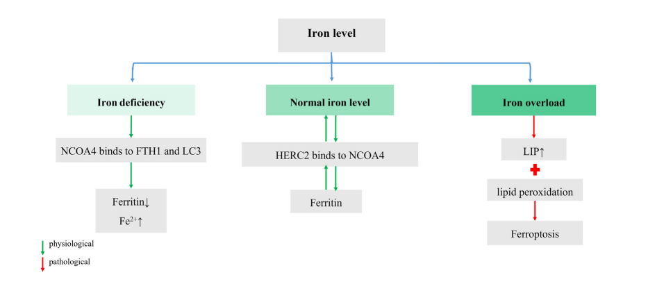
- NCOA4 is an iron-sensing protein whose levels are regulated by intracellular iron status:
- When iron levels are high, NCOA4 abundance is low (it is ubiquitinated by ubiquitin ligase HERC2), thereby promoting ferritin accumulation and iron capture.
- When intracellular iron levels are low, NCOA4 levels increase, resulting in ferritinophagy-mediated degradation of ferritin with release of ferric ions (Fe3+) and their subsequent conversion into ferrous ions (Fe2+).
- NCOA4-deficient cells in vitro fail to activate ferritinophagy and are associated with decreased bioavailable iron.9
- NCOA4-deficient mice demonstrate:10
- Iron accumulation in the liver and spleen
- Increased levels of:
- Transferrin saturation
- Serum ferritin
- Liver hepcidin
- Decreased levels of duodenal ferroportin
- Mild microcytic hypochromic anemia
- In summary:
- Ferritinophagy is initiated by binding of NCOA4 to ferritin.
- NCOA4 is a selective cargo receptor for autophagic turnover of ferritin.
- NCOA4 acts as an iron sensor that is:
- Degraded in condition of iron excess
- Binds ferritin for the recycling of iron under conditions of iron starvation