Schistocytes

What are schistocytes?

  • Schistocytes are circulating fragments of red blood cells or RBCs from which cytoplasmic fragments have been lost, suggestive of:
    • Thrombotic microangiopathy
    • Malfunctioning prosthetic valve
  • Schistocytes are always smaller than intact red cells and may be recognized as:
    • Triangles
    • Small crescents
    • Helmet cells
    • Keratocytes (horn cells)
    • Microspherocytes
  • Schistocytes are homogeneously stained in most cases with the exception of keratocytes, which may have some central pallor.
  • RBC fragments similar to schistocytes can be found in non-TMA-related genetic or acquired RBC disorders:
    • Including:
      • Red blood cell membrane defects
      • Thalassemia
      • Megaloblastic anemia
      • Primary myelofibrosis
      • Thermal injuries
    • Typically show highly variable shapes and are associated with marked anisopoikilocytosis and a wide range of additional RBC size and morphological changes.
  • Schistocytes are detected in the peripheral blood smear stained using standard procedures and observed by light microscopy, and expressed as a percentage of RBCs.
  • Precise morphological criteria for the recognition and enumeration of schistocytes still remain poorly defined, leading to observer bias.
  • A schistocyte count should be considered clinically meaningful if they represent the main morphological red blood cells abnormality in the smear (other than signs of erythropoietic regeneration).
  • The univocal mechanism of schistocyte formation is mechanical damage to the membrane caused by fibrin strands on the endothelial surface and/or excess of turbulence of blood. They develop because red blood cells are sheared as they try to pass through small vessels narrowed by microthrombi.

Reference

The following are examples of 4 of the 5 schistocyte types recognized by the Schistocyte Working Group of the International Council for Standardization in Haematology (ICSH):

The presence of schistocytes helps to narrow the differential diagnosis to a thrombotic microangiopathy (TMA), including thrombotic thrombocytopenia purpura (TTP), hemolytic uremic syndrome (HUS) or disseminated intravascular coagulation (DIC).

Can the number/frequency of schistocytes help make a diagnosis of TMA?

  • Schistocyte count is up to 0.2% in normal individuals.
  • In DIC, the schistocyte count is typically between 0.2% and 1%. However, counts can exceed 1%.
  • In TTP and HUS:
    • A robust morphological indication for the diagnosis of thrombotic microangiopathic anemia in adults should be recognized when the percentage of schistocytes is above 1%. However, a lack of schistocytes does not exclude a priori the diagnosis of TMA (learn more here). There is no threshold schistocyte number below which the possibility of TTP can be excluded.
    • In the appropriate clinical context, more than two schistocytes per high-powered field or more than 1.0% schistocytes on a blood film is considered pathologic; however, these cut-offs have not been validated (learn more here).

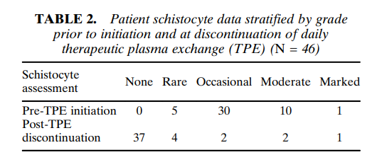
The following are results from a a cohort of 46 patients with TTP and severe ADAMTS13 deficiency. Note the wide variation in schistocyte counts:

Schistocyte burden is reported as rare (<1%), occasional (1–5%), moderate (6–25%), or marked (>25%)

Bottom line:

A high number of schistocytes on the peripheral smear suggests a diagnosis of thrombotic microangiopathy (TMA), but cannot reliably distinguish between TTP and other types of TMA.

Prev
 1 / 0 
Next