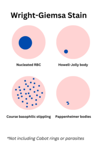
Red Blood Cell Inclusions
Introduction
- Red blood cell (RBC) inclusions include:
- Those visible by Wright-Giemsa staining:
- Nucleated RBC
- Howell–Jolly bodies (nuclear fragments)
- Pappenheimer bodies (iron-containing autophagosomes)
- Cabot rings (mitotic spindle remnants)
- Basophilic stippling (aggregates of ribosomes)
- Hb C crystals
- Parasites
- Those visible by other stains:
- Iron stains:
- Siderosomes (iron-containing inclusions)
- Confirmation of Pappenheimer bodies
- Intravital stains:
- Heinz bodies (denatured globin)
- HbH inclusions
- Iron stains:
- Most RBC inclusions are found in young RBCs (reticulocytes).
- The inclusions represent nuclear or cytoplasmic remnants.
- They must be distinguished from:
- Stain precipitate
- Bubbles
- Dirt on slide
- Overlying platelets
- Those visible by Wright-Giemsa staining:
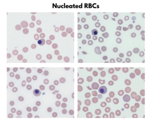
Nucleated red blood cells
- Represents the presence of normoblasts in the peripheral blood, typically at orthochromatophilic stage of maturation.
- Typically enumerated and reported as number of nucleated red blood cells (nRBCs) per 100 white blood cells.
- Not necessary to distinguish the exact stage of maturation in the peripheral blood. The term nRBC encompasses all normoblasts circulating in the peripheral blood, regardless of maturation stage.
- Compared with a lymphocyte, the nRBC has:
- Pinkish cytoplasmic color
- Lower N:C ratio
- Dark purple chromatic color
- Coarse condensation of chromatin (pyknotic)
- Physiologically present at birth, generally disappear within 3-5 days.
- May be seen in:
- Bone marrow replacement processes including:
- Metastatic tumor
- Marrow infiltration with leukemia/lymphoma
- Marrow fibrosis
- Marrow granulomas
- Rapid RBC production, for example hemolytic anemias
- Bone marrow replacement processes including:
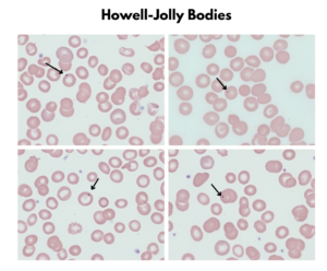
Howell-Jolly Bodies
- Spherical, dark purple with Wright’s stain, vary widely in size and usually eccentrically located in the cell.
- Compared with Pappenheimer body:
- Larger
- Rounder
- Do not form tight clusters in pairs or tetrads
- Nuclear remnants or the result of abnormal mitosis in which a single chromosome fails to participate in the interphase nucleus and becomes detached:
- The likelihood of the latter happening increases if the nucleus contains > 4N number of chromosomes prior to cell division as occurs in megaloblastic anemia.
- Normally the spleen removes any Howell-Jolly body, hence their presence in the setting of asplenia.
- Seen in:
- Hyposplenism
- Asplenism
- Severe hemolytic anemia
- Megaloblastic anemia
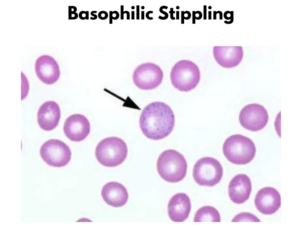
Basophilic Stippling
- Consists of multiple, uniform, evenly distributed fine or coarse dark dots (granules) scattered within the cytoplasm of RBCs.
- Two types of basophilic stippling:
- Course:
- RBC has variably sized (up to large) basophilic ‘granular’ discolorations across its entire cytoplasm on a Wright-stained film.
- Never a normal finding; suggests impaired hemoglobin synthesis.
- Seen in:
- Thalassemia
- Lead poisoning
- Myelodysplastic syndrome
- Sideroblastic anemia
- Congenital dyserythropoietic anemia
- Fine:
- RBC has small, uniform, punctate basophilic dots across its entire cytoplasm, on a Wright-stained film.
- Associated with reticulocytosis; usually found in red cells that are larger and more polychromatophilic (purple) compared with neighboring non-stippled RBCs.
- Of no clinical consequence.
- Course:
- Stippled cells are created when reticulocytes from patients with incomplete RNA degeneration (or abnormal ribosomes) are dried slowly or stained supravitally with new methylene blue:
- The RNA-containing ribosomes of reticulocytes aggregate during these processes and thus become visible as stippling.
- In lead poisoning and thalassemia, the altered reticulocyte ribosomes have a greater propensity to aggregate, forming larger granules (coarse stippling).
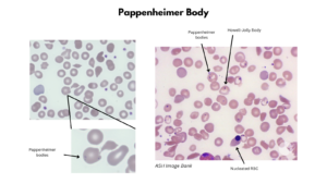
Pappenheimer bodies
- Pappenheimer bodies, or siderotic granules, appear in Wright-stained preparations as small, irregular basophilic (purple) coccoid granules < 1 um in diameter, sometimes clustered, and generally located near the cell periphery.
- Compared with basophilic stippling, Pappenheimer bodies occupy only one portion or region of the RBC.
- Compared with Howell-Jolly bodies, Pappenheimer bodies are:
- Smaller
- Less round, more angular
- Form tight clusters in pairs or tetrads
- Wright Giemsa stains the protein matrix of the granules, while Prussian blue stains the nonheme iron component.
- Cells containing Pappenheimer bodies are termed siderocytes.
- Created when autophagosomes in RBCs digest abnormal iron-containing mitochondria.
- The autophagosomes are normally discharged from the cytoplasm or removed by the pitting action of the spleen.
- Seen in anemias which have in common a defect of incorporation of iron into the hemoglobin molecule.
- Observed in:
- Asplenia
- Megaloblastic anemia
- Thalassemia
- Hemolytic anemia
- Sideroblastic anemia
- Congenital dyserythropoietic anemia
Cabot Rings
- Appear as thread-like red or purple loops which may be single, double, or twisted as a figure-of-eight:
- Seen in:
- Megaloblastic anemia
- Severe anemia
- Leukemia
- Lead poisoning
- Other causes of dyserythropoiesis
| Inclusion | Composition | Appearance on WG stain | Clinical Conditions |
|---|---|---|---|
| nRBC | DNA | Single, dark purple nucleus, agranular cytoplasm with varying degrees pf hemoglobinization (pinkess) | Newborn. severe stress reaction, myelofibrosis; thalassemia, hemolytic anemia, MDS |
| Howell-Jolly body | DNA | Round blue granules, 1 um diameter, at cell periphery, usually single, may be multiple | Hypo/asplenia, severe hemolytic anemia, megaloblastic anemia |
| Basophilic stippling (coarse) | Precipitate of ribosomes | Punctate blue granules | Thalassemia, lead poisoning, MDS, sideroblastic anemia, congenital dyserythropoietic anemia |
| Pappenheimer bodies | Iron-containing autophagosome | Blue-purple granules, < 1 um diameter, at cell periphery; may form doublets, iron stain positive, DNA stain negative | Asplenia, megaloblastic anemia, thalassemia, hemolytic anemias, congenital dyserythropoietic anemia |
| Cabot rings | Remnants of mitotic spindle | Red-purple thread-like rings | Megaloblastic anemia, severe anemia, leukemia, lead poisoning, other causes of dyserythropoiesis |
| Parasites | Various parasites | Variable appearance | Malaria, babesiosis |
References