Postscript
A metaphor is a figure of speech whereby a word or phrase ordinarily used in one domain (source domain) is applied to another (target domain) for the purpose of indicating a resemblance between them. Key to the definition is that its literal meaning is bypassed as it takes on a new figurative meaning. Metaphors are not only rhetorical or poetic devices. They can be used as cognitive tools to provide insights into how things are. Metaphors play a part in generating hypotheses, extending explanation, and stimulating original thought. Simply put, our understanding of the world is derived from metaphors. Metaphors are used in everyday use of language, and not only in language but in thought and action as well. In medicine, they are particularly useful for communicating complex ideas, both between physicians and between physicians and their patients. Consider, for example, the ubquiqitous use of war metaphors (body as battlefield):
- The fight against cancer
- The war on cancer
- Her patient has given up the fight
- He lost the battle to cancer
- Cells battle for supremacy and survival
- Lymphocytes are deployed or mobilized in infection
- Platelets are released into the circulation with their formidable payload
- Innate immunity is the first line of defense
- Treatment may cause collateral damage
- This drug is a magic bullet
To see these types of metaphors in action, consider the following passage from River of Life (the book title itself being a nice example of metaphor):
The blood “Stands as a constant bulwark against invasion by the swarms of microbes that infest our environment… [without the defenses provided by our blood], we probably could not survive a single day against the assaults of our microscopic foes…. Unlike the phagocytes which are non-discriminatory soldiers attacking any foreign substance that invades the body tissue, the antibodies are active against only a single type of invader.”
Patients also used metaphors to describe their personal experience of illness:
- A dark cloud hangs over me
- The nausea comes in waves
- It feels as if there is an elephant sitting on my chest
Metaphors require that the listener or reader compares the source and its target domains, a process termed mapping. Source and target domains can be analyzed in terms of their semantic nearness. If the source and target are from similar fields, the metaphor is termed local. If they are from different fields, the metaphor is referred to as distant. Distant metaphors are also characterized as possessing tension. The more tension that exists between source and target domains the harder we have to work at drawing the appropriate connections.
The lifespan (or career) of a metaphor varies widely. Some are used in an ad hoc fashion, never to be used again. Others become so widely used that they are no longer considered metaphors (they are termed dead metaphors). In some cases, the literal interpretation of the word expands to include the former metaphorical meaning. For example, consider the Webster Dictionary definition of sickle cell:

The term sickle was introduced by James Herrick when he first described a patient with sickle cell disease in 1910. As a newly introduced term, it would would have provoked some thought as listeners and readers mapped the source domain (an agricultural tool) with a bizarrely shaped red blood cell. But the term became so commonly used over the next 100 years that it took on a literal meaning.
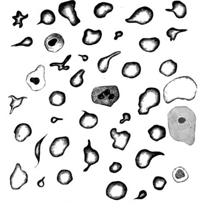
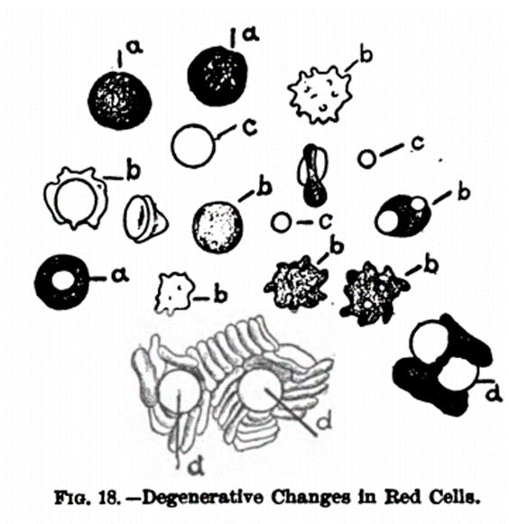
And this brings us to focus of the tweet: poikilocytes. Imagine being the first to look down the eyepiece of a microscope and observing red blood cells from patients that had odd shapes. Here are two examples from the turn of the 20th century:


Early investigators simply lumped all abnormally shaped red blood cells together under the umbrella term poikilocyte. Eventually, efforts were made to differentiate between different types of poikilocytes in an effort to link a specific poikilocyte types to specific diseases. For example, DeCosta wrote in 1910:

There are two points to make about this passage. First, note that DeCosta is careful to point out that the red cell resembles the shape of a gourd or a horseshoe. This is a simile, not a metaphor. A simile overtly indicates its figurative sense to a reader by using words “like”, “resembles” or “as” to compare the two things. Second, metaphors are highly context dependent and their usefulness requires some familiarity on the part of the listener/reader about the source domain. For example, many of us would be hard pressed to know what a gourd looks like, so it has outlived its usefulness as a metaphor. Here are additional similes that have not survived to this day:

Even by 1950, there was no uniform description of various poikilocytes:
In case you didn’t know what belaying pins were!

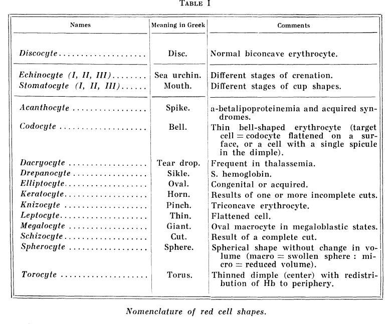
By 1970, there was no beating around the bush. Red cells were no longer “shaped” like teardrops. They were teardrops (or more accurately dacrocytes at the time) In 1972, Bessis appraised the status of poikilocytes:
The various shapes that the red cell can assume are just beginning to be analysed in a critical fashion. It is now possible to assign names to most of the cells which have been anonymously designated as poikilocytes. Thus, the erythrocyte may become the first living cell permitting us to assess its internal molecular structure on the basis of its external configuration.

The metaphorical nature of poikilocyte terminology extends beyond mere shape comparisons. Take the sickle cell as an example. The harvesting tool has a blade made of steel. Like the sickle cell, it is stiff and inflexible. The teardrop conjures images of motion and sadness, just as teardrop cells – circulating in the blood – often indicate serious underlying disease.
Are there other red cell shapes that remain to be discovered in disease? You might think we have seen it all (just as I am sure some investigators in the early 1900s assumed they had captured all possible poikilocyte types). But consider a study from 2018 that described a new poikilocyte:

These cells were shown to be present in significant numbers in patients with hereditary elliptocytosis, iron deficiency, and B12 and folate deficiency. Note how the authors describe the cells as “fish shaped”. This is really more simile than metaphor: the cells are similar to a fish in their shape. If the findings prove to be clinically relevant, we will likely see a day when the cells are simply called fish cells.
Indeed, one of the replies to our tweet already made that jump:

Over time, with repetitive use, any cross-domain mapping between a fish and a red cell may fade, and the metaphor may ultimately die. Who knows, maybe the Webster Dictionary definition of fish may include the fish cell some day! OK, maybe not 🙂
- 15,850 patients examined in the vascular laboratory
- 2568 (16.2%) had DVT
- Thrombosis in unusual sites occurred in 14 patients giving a prevalence of 0.088% among all patients or 0.54% in patients with DVT
- 8 cases of deep femoral vein thromboses
- 2 thromboses in the femoropopliteal vein
- 1 in the deep external pudendal vein
- 1 in a muscular thigh vein
- 2 patients with thromboses in the lateral thigh vein
The veins examined were the deep external pudendal
vein which is seen uniting the common femoral vein opposite to saphenofemoral junction
The internal pudendal veins are the set of accompanying veins to the internal pudendal artery draining the perineal region to empty into the internal iliac vein.
The internal pudendal vein drains oxygen-depleted blood from the perineum, which is the area between the exterior genitals and anus, and the external genitalia. The drained region includes the bulb of the penis (in males) or clitoris (in females), the anal region, and the urogenital region. Tributaries of the internal pudendal vein include the vein of the bulb (in males), the posterior labial vein (in females), the scrotal vein (in males), and the inferior rectal vein. The internal pudendal vein drains into the internal iliac vein. Despite its location, the deep dorsal vein, which drains the erectile bodies of the penis (in men), does not pass into the internal pudendal vein.
The internal pudendal veins (internal pudic veins) are a set of veins in the pelvis. They are the venae comitantes of the internal pudendal artery.
They begin in the deep veins of the vulva and of the penis, issuing from the bulb of the vestibule and the bulb of the penis, respectively. They accompany the internal pudendal artery, and unite to form a single vessel, which ends in the internal iliac vein.
They receive the veins from the urethral bulb, the perineal and inferior hemorrhoidal veins.
The deep dorsal vein of the penis communicates with the internal pudendal veins, but ends mainly in the pudendal plexus.

