
When a Bleeding Disorder Causes a Deep Hemorrhage

When a Bleeding Disorder Causes a Deep Hemorrhage
Managing a large chest-wall hematoma in type 2B von Willebrand disease

By William C. Aird, MD

Case Presentation

A 50-year-old woman with a historical diagnosis of type 2B von Willebrand disease (VWD) presents with four days of progressively worsening right posterior chest and back pain.

She recalls no major trauma, although she had recently reached overhead, walked a large dog that occasionally pulled on its leash, and underwent a deep tissue massage after the pain began.

Because of severe pain, she presents to an outside emergency department.

CT angiography demonstrates a 16-cm hematoma involving the right chest wall near the serratus anterior muscle with active contrast extravasation.
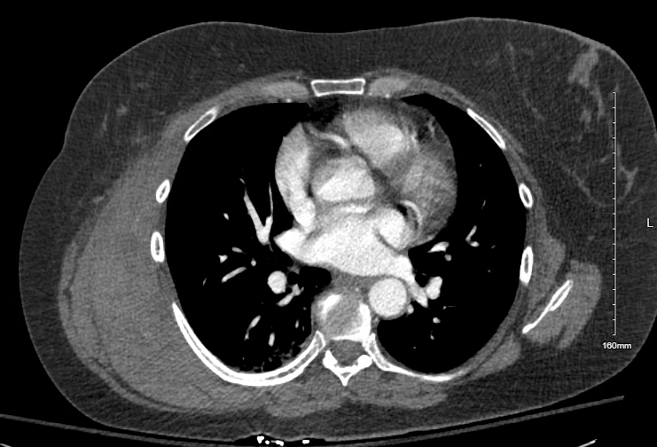
“Chest wall: There is a right posterolateral hematoma of the chest wall overlying the rib cage and deep to the serratus anterior muscle tear. The hematoma extends up to the second rib and inferiorly down to the level of the 10th rib. It measures about 16 x 16 x 5 cm. There are multiple punctate foci of contrast density within the hematoma consistent with active extravasation.”

Initial laboratory studies show:

TestResult
Hemoglobin10.5 g/dL
Platelet count300 ×10⁹/L
PTNormal
aPTTNormal

She receives tranexamic acid and desmopressin before transfer to a tertiary care center.

Despite the imaging findings, she remains hemodynamically stable.

Bleeding History

She has experienced lifelong easy bruising and heavy menstrual bleeding.

During delivery of twins she required desmopressin after postpartum hemorrhage.

All three of her daughters have also been diagnosed with type 2B VWD.

She has never previously experienced a major spontaneous soft-tissue hemorrhage.

Which feature of this presentation is most concerning?

a
Normal platelet count
b
Normal PT and aPTT
c
Large chest-wall hematoma with active contrast extravasation
Large intramuscular or chest-wall hematomas are uncommon in VWD but may occur. The combination of: a large expanding hematoma active contrast extravasation declining hemoglobin suggests clinically important bleeding requiring close monitoring and targeted hemostatic therapy. Although PT and aPTT are normal, normal coagulation screening tests do not exclude clinically significant VWD.
d
History of easy bruising

Explanation

Large intramuscular or chest-wall hematomas are uncommon in VWD but may occur.

The combination of:

  • a large expanding hematoma
  • active contrast extravasation
  • declining hemoglobin

suggests clinically important bleeding requiring close monitoring and targeted hemostatic therapy.

Although PT and aPTT are normal, normal coagulation screening tests do not exclude clinically significant VWD.

Diagnostic Background

Historical evaluation had established a diagnosis of type 2B VWD.

Laboratory studies obtained during this admission demonstrated:

TestResult
VWF antigen90 IU/dL
Platelet-dependent VWF activity37 IU/dL
FVIII activity69 IU/dL

The ratio of platelet-dependent VWF activity to VWF antigen is markedly reduced, consistent with qualitative VWF dysfunction.

These studies support a type 2 VWD phenotype but do not independently establish the subtype.

Which laboratory finding is most consistent with a qualitative defect in von Willebrand factor?

a
Low VWF antigen with proportionately reduced activity
b
Reduced platelet-dependent VWF activity despite near-normal VWF antigen
Patients with type 2 VWD often have: relatively preserved VWF antigen disproportionately reduced platelet-dependent VWF activity The activity-to-antigen ratio therefore falls below normal, reflecting impaired VWF function rather than decreased quantity.
c
Isolated prolongation of PT
d
Severe thrombocytopenia

Explanation

Patients with type 2 VWD often have:

  • relatively preserved VWF antigen
  • disproportionately reduced platelet-dependent VWF activity

The activity-to-antigen ratio therefore falls below normal, reflecting impaired VWF function rather than decreased quantity.

Initial Management

Interventional radiology reviews the CT scan.

Although active contrast extravasation is present, the patient remains:

  • normotensive
  • without tachycardia
  • clinically stable

Compression is applied over the hematoma, and serial hemoglobin measurements are obtained.

Hemoglobin falls further:

TimeHemoglobin
Admission10.5 g/dL
Later that day9.2 g/dL
Hospital Day 29.0 g/dL

Because of continued bleeding, she receives 6,000 units of Humate-P, a plasma-derived concentrate containing both VWF and factor VIII.

Why is desmopressin generally avoided in patients with type 2B VWD who develop major bleeding?

a
It may worsen abnormal platelet-VWF interactions and precipitate thrombocytopenia
Desmopressin releases endogenous VWF from endothelial storage sites. In type 2B VWD, the released VWF has increased affinity for platelet glycoprotein Ib. This may: enhance platelet binding precipitate thrombocytopenia provide less effective hemostasis than VWF replacement For major bleeding, current practice generally favors VWF-containing concentrate rather than desmopressin.
b
It rapidly consumes factor VIII
c
It increases fibrinolysis
d
It inhibits endogenous VWF production

Explanation

Desmopressin releases endogenous VWF from endothelial storage sites.

In type 2B VWD, the released VWF has increased affinity for platelet glycoprotein Ib.

This may:

  • enhance platelet binding
  • precipitate thrombocytopenia
  • provide less effective hemostasis than VWF replacement

For major bleeding, current practice generally favors VWF-containing concentrate rather than desmopressin.

Clinical Course

Following administration of Humate-P, the patient’s hemoglobin stabilizes.

Serial examinations demonstrate no further expansion of the hematoma.

Because she remains clinically stable, interventional radiology determines that angiographic embolization is unnecessary.

She is discharged home with hematology follow-up and repeat CBC monitoring.

Which factor most strongly supported continued conservative management rather than immediate embolization?

a
The diagnosis of VWD
In type 2B VWD, the released VWF has increased affinity for platelet glycoprotein Ib. This may: enhance platelet binding precipitate thrombocytopenia provide less effective hemostasis than VWF replacement For major bleeding, current practice generally favors VWF-containing concentrate rather than desmopressin.
b
Normal PT and aPTT
c
Hemodynamic stability with stabilization after hemostatic therapy
Active contrast extravasation does not automatically require embolization. Management should integrate: hemodynamic status hemoglobin trajectory clinical examination response to hemostatic therapy feasibility of local intervention In this patient, stabilization after VWF replacement and compression allowed successful nonoperative management.
d
Normal platelet count

Explanation

Active contrast extravasation does not automatically require embolization.

Management should integrate:

  • hemodynamic status
  • hemoglobin trajectory
  • clinical examination
  • response to hemostatic therapy
  • feasibility of local intervention

In this patient, stabilization after VWF replacement and compression allowed successful nonoperative management.

Teaching Points

  • Type 2B VWD is a qualitative disorder of VWF function rather than VWF quantity.
  • Normal PT, aPTT, and even a normal platelet count do not exclude clinically significant type 2B VWD
  • A reduced platelet-dependent VWF activity relative to VWF antigen suggests qualitative VWF dysfunction.
  • For major bleeding in type 2B VWD, VWF-containing concentrate is generally preferred over desmopressin.
  • Management of major bleeding requires integration of clinical status, laboratory trends, imaging findings, and disease-specific hemostatic therapy.

Why This Case Matters

Von Willebrand disease is often thought of as a disorder of mucosal bleeding, but major soft-tissue hemorrhage can occur and requires a different level of clinical urgency.

This case illustrates three important principles. First, laboratory interpretation depends on assessing VWF function as well as quantity. Second, identifying the specific VWD subtype influences treatment decisions, particularly the choice between desmopressin and VWF replacement. Finally, successful management of major bleeding requires more than interpreting imaging findings. Decisions about intervention depend on the patient’s overall clinical trajectory, emphasizing that treatment is guided by the patient, not the CT scan.

Prev
 1 / 16 
Next