
Can severe inflammation conceal absolute iron deficiency even when ferritin exceeds 300 ng/mL?
A Case That Challenges a Clinical Rule
I have long used a practical rule when interpreting iron studies: a serum ferritin below 20 ng/mL establishes absolute iron deficiency, whereas a ferritin above 100 ng/mL virtually always excludes it, even in the presence of inflammation. Important exceptions include ferritin release associated with cellular injury and rare inherited hyperferritinemia syndromes, in which serum ferritin may be elevated independently of iron stores. Although diagnostic thresholds vary among guidelines and clinical settings, this rule has generally served me well.
Recently, a patient challenged that assumption.
A young woman was hospitalized with a severe multisystem inflammatory illness characterized by high fevers, markedly elevated inflammatory markers, transient anemia, and hepatocellular injury. Despite an extensive evaluation, no definitive diagnosis was established. She gradually recovered.
During hospitalization, her ferritin was 354 ng/mL. Approximately five weeks later, it had fallen to 20 ng/mL.
The accompanying laboratory changes were equally striking.
| Laboratory measurement | Acute illness | Recovery (~5 weeks later) |
|---|---|---|
| Ferritin (ng/mL) | 354 | 20 |
| CRP (mg/L) | >300 | 19.9 |
| Serum iron (µg/dL) | 7 | 40 |
| Transferrin (mg/dL) | 141 | 256 |
| Transferrin saturation (%) | 4 | 12 |
| Hemoglobin (g/dL) | 10.2 | 14.2 |
During hospitalization, AST reached approximately 268 U/L and ALT approximately 106 U/L, consistent with hepatic involvement in the systemic illness. There was no documented major bleeding to explain rapid depletion of iron stores.
The ferritin concentration fell by more than 90% in just over a month, reaching 20 ng/mL, a value strongly indicative of low iron stores. This decline occurred alongside substantial improvement in systemic inflammation, although CRP remained elevated at the later measurement.1
This raises the central question: Was the patient already absolutely iron deficient when her ferritin was 354 ng/mL, with severe inflammation temporarily obscuring depleted iron stores? If so, this would represent a striking exception to my longstanding rule. What biological mechanisms could account for such a marked ferritin elevation despite depleted iron stores?
What Does Serum Ferritin Actually Measure?
Ferritin is an intracellular iron-storage protein composed of 24 subunits of two types, heavy (H) and light (L), that assemble into a hollow shell capable of storing thousands of iron atoms. By incorporating iron into a relatively nonreactive mineral core, ferritin protects cells from the potentially toxic effects of unbound iron while maintaining an accessible intracellular reserve.
Under relatively steady-state conditions, serum ferritin correlates with body iron stores. This relationship makes it one of the most useful laboratory tests for diagnosing absolute iron deficiency.2
Yet circulating ferritin differs substantially from its intracellular counterpart. In a study characterizing serum ferritin in mice, Cohen and colleagues demonstrated that circulating ferritin consisted predominantly of L-subunits, contained relatively few H-subunits, and was iron-poor compared with intracellular storage ferritin.3 They also identified a truncated L-subunit consistent with processing through a lysosomal pathway.
Serum ferritin therefore does not directly measure the iron contained within intracellular ferritin stores. Instead, the assay measures the concentration of a circulating, largely iron-poor protein. Although macrophages are an important source of serum ferritin, the physiological purpose of its secretion remains uncertain.
It is remarkable that the concentration of this iron-poor circulating protein correlates as well as it does with total-body iron stores. However, the relationship can become distorted during inflammation or tissue injury, when ferritin production, secretion, and release from damaged cells may change independently of iron stores. Ferritin released through cellular injury may also differ in its iron content from ferritin that is actively secreted.
Several processes can influence serum ferritin concentrations:
- Intracellular iron availability and ferritin synthesis.
- Inflammatory regulation of ferritin gene expression and translation.
- Regulated secretion of ferritin from cells.
- Release of ferritin associated with tissue injury.
- Changes in ferritin degradation and clearance.
To understand why serum ferritin generally falls with iron depletion, and why this relationship may become distorted during inflammation, we must first examine how ferritin synthesis is regulated.
How Iron Deficiency Regulates Ferritin Synthesis
Ferritin synthesis is regulated through the iron-responsive element/iron regulatory protein (IRE/IRP) system.
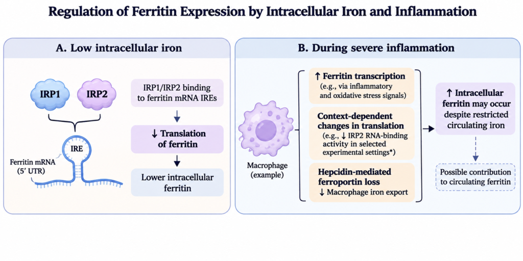
Ferritin H- and L-chain messenger RNAs contain an IRE in their 5′ untranslated regions. When intracellular iron availability is low, IRP1 and IRP2 bind these elements and inhibit translation. Ferritin protein synthesis is therefore reduced even when ferritin mRNA is present.
When intracellular iron availability increases, IRP binding is diminished, allowing translation to proceed.
The two IRPs are not functionally identical. IRP1 can exist either as an RNA-binding protein or as a cytosolic aconitase containing an iron-sulfur cluster. Under many physiological conditions, IRP1 predominantly occupies its aconitase form, whereas IRP2 makes an important contribution to iron-responsive RNA binding and translational regulation.4
The IRE/IRP system also regulates other proteins involved in iron metabolism, sometimes in the opposite direction. For example, transferrin receptor 1 (TfR1) mRNA contains IREs in its 3′ untranslated region. When intracellular iron is scarce, IRP binding stabilizes TfR1 mRNA, increasing receptor expression and the cell’s capacity to acquire transferrin-bound iron. Thus, the same regulatory system decreases ferritin synthesis while increasing TfR1 expression, allowing cells to conserve iron and increase its uptake.
This regulatory system provides a mechanistic explanation for the low ferritin concentrations observed in absolute iron deficiency. When intracellular iron is scarce, ferritin translation is suppressed, reducing intracellular ferritin production and presumably the amount available for secretion into the circulation.
Inflammation introduces another layer of regulation. Cytokines and oxidative stress can increase ferritin gene transcription through regulatory pathways involving NF-κB and antioxidant-response elements. These signals increase ferritin mRNA abundance, while the IRE/IRP system continues to regulate its translation.5
My longstanding assumption has been that, in profound absolute iron deficiency, iron-dependent translational repression generally dominates over inflammation-driven transcriptional activation. Increasing ferritin mRNA abundance should have limited effect if the resulting transcripts cannot be efficiently translated.
However, this assumes that inflammatory signaling does not substantially modify iron-dependent translational repression and that there is no additional release of ferritin from injured cells. Neither assumption is necessarily correct.
Inflammation Can Modify Ferritin Translation
As discussed above, inflammatory and oxidative signals can increase ferritin gene transcription. However, their effects are not confined to transcription. Experimental studies demonstrate that these signals can also modify IRP activity and ferritin translation, with effects that depend on the stimulus, timing, and intracellular iron availability.
Tsuji and colleagues demonstrated that oxidative stress initially increased IRP1 RNA-binding activity, favoring translational repression. Subsequently, IRP1 activity returned toward baseline, permitting translation of newly synthesized ferritin transcripts and increasing ferritin protein production.6
In a different experimental model, Recalcati and colleagues found that stimulation of murine macrophages with interferon-γ and lipopolysaccharide slightly increased IRP1 activity but markedly decreased IRP2 activity, accompanied by increased ferritin synthesis.7 Importantly, iron chelation prevented the reduction in IRP2 activity, suggesting that the response depended at least partly on intracellular iron availability.
The investigators suggested that inflammatory signaling might act indirectly by increasing intracellular iron availability, which in turn reduces IRP2 activity. This interpretation is consistent with the observation that iron chelation prevented the response, but it does not establish that this is the only mechanism involved.
These findings indicate that inflammatory and oxidative signals can modify ferritin translational regulation in different directions. They do not demonstrate that inflammation can overcome iron-dependent translational repression when intracellular iron is profoundly depleted.
The results therefore qualify, rather than simply invalidate, the hypothesis that translational repression generally dominates in absolute iron deficiency.
These experimental findings raise the possibility that inflammatory and oxidative signals can counteract the translational repression of ferritin synthesis associated with iron deficiency. However, they do not establish that this occurs when intracellular iron is profoundly depleted, or how large an increase in serum ferritin might result. Clinical studies comparing serum ferritin with bone marrow iron provide a way to examine the latter question.
How High Can Ferritin Rise in Absolute Iron Deficiency?
Studies comparing serum ferritin with stainable bone marrow iron provide an empirical basis for estimating how high ferritin can rise in patients with absolute iron deficiency.
In a 2020 ASH Education Program review, Fertrin summarized a systematic review of 38 studies involving 1,023 patients with absent stainable bone marrow iron. The overall mean ferritin concentration was 82.4 ng/mL, although mean values varied substantially across disease groups, ranging from 33.6 ng/mL in patients with rheumatoid arthritis to 158.3 ng/mL in patients with hematologic disorders. These figures represent group means rather than maximum individual values. Outside chronic kidney disease and certain heart failure populations, absent marrow iron was rarely associated with ferritin concentrations above approximately 200 ng/mL.8
The most striking departures from this pattern occurred in dialysis-dependent chronic kidney disease. In these patients, absent marrow iron was sometimes documented despite ferritin concentrations exceeding 300 ng/mL and occasionally 1,000 ng/mL. However, these patients have complex disturbances of iron metabolism, often involving dialysis-related blood loss, intravenous iron treatment, and erythropoiesis-stimulating agents. Their findings cannot be directly extrapolated to otherwise healthy individuals experiencing an acute inflammatory illness.9
The 2021 British Society of Gastroenterology (BSG) guideline provides a complementary clinical perspective. It states that absolute iron deficiency is unlikely when serum ferritin exceeds 150 ng/mL, even in the presence of inflammation.10
These two sources approach the question differently. Fertrin summarizes empirical observations using bone marrow iron as the reference standard, whereas the BSG guideline translates the available evidence into a practical diagnostic statement. Neither establishes an absolute exclusion threshold.
Taken together, the findings support the usefulness of a relatively high ferritin concentration as evidence against absolute iron deficiency. They also suggest that my longstanding threshold of 100 ng/mL may have been more conservative than necessary. A threshold of 150 ng/mL, or perhaps approximately 200 ng/mL in selected clinical contexts, has support in the published literature, although these values should be understood as probabilistic rather than absolute boundaries.
The patient’s ferritin of 354 ng/mL exceeds all three thresholds. If she was already absolutely iron deficient during hospitalization, the result would represent an unusual exception to the expected relationship between serum ferritin and iron stores.
The published cases of high ferritin with absent marrow iron establish that such coexistence is possible. They do not establish which mechanisms are responsible or whether those mechanisms explain this patient’s findings.
Macrophage Iron Retention During Inflammation
Inflammation changes the distribution of iron within the body. IL-6 stimulates hepatic hepcidin production, which promotes ferroportin internalization and degradation. Iron export from macrophages is consequently restricted, producing the familiar combination of hypoferremia, iron-restricted erythropoiesis, and macrophage iron sequestration seen in anemia of inflammation.
This mechanism explains why circulating iron may be profoundly reduced while iron remains present within macrophages. With superimposed absolute iron deficiency, however, macrophage iron stores would also tend to become depleted.
Macrophages continue to recycle iron from senescent erythrocytes during absolute iron deficiency, but the amount they retain depends on the balance between inflammatory hepcidin stimulation and the hepcidin suppression normally associated with iron deficiency.
Moreover, total macrophage iron content is not equivalent to the intracellular regulatory iron pool controlling IRP activity. Iron retained within ferritin or other storage compartments may not be immediately available to the iron-sensing machinery.
Consequently, classical macrophage iron sequestration provides a well-established explanation for elevated ferritin in anemia of inflammation, but it does not necessarily explain substantial ferritin elevations in patients with absolute iron deficiency.
It is uncertain how much macrophage iron retention and altered translational regulation can contribute to circulating ferritin when storage iron is depleted. These mechanisms might explain a modest inflammatory increase, but whether they can independently account for concentrations above 300 ng/mL remains unresolved.
Macrophage Ferritin Secretion
Changes in intracellular ferritin abundance can be reflected in circulating ferritin concentrations through regulated secretion. This provides an important link between the cellular mechanisms discussed above and the serum ferritin concentrations measured in clinical practice.
In their 2010 study, Cohen and colleagues provided evidence that macrophages are major contributors to circulating ferritin in mice. They also cited earlier genetic studies showing that total or macrophage-specific deletion of IRP2 was associated with elevated serum ferritin, whereas hepatocyte-specific or intestinal epithelial cell-specific deletion did not produce the same response.11
These observations complement the Recalcati findings and support a relationship between macrophage IRP2 regulation, ferritin synthesis, and circulating ferritin concentrations.
Cohen and colleagues also provided evidence for a nonclassical secretory pathway involving lysosomal trafficking. Ferritin can therefore enter the circulation through secretion from intact macrophages without requiring cellular injury or destruction.
Although intracellular ferritin abundance and circulating ferritin concentrations are biologically linked, secretion is itself regulated. Experimental studies of vesicular trafficking have demonstrated that serum ferritin concentrations can change without corresponding changes in conventional markers of systemic iron status.12
Thus, increased intracellular ferritin synthesis or accumulation may be accompanied by increased secretion, but the relationship is not necessarily proportional. The central question in absolute iron deficiency remains whether severe inflammation can increase intracellular ferritin sufficiently to produce a substantial rise in serum ferritin despite iron-dependent translational repression.
Ferritin Degradation and Ferritinophagy
Intracellular ferritin abundance depends on both synthesis and degradation.
Ferritinophagy is a selective autophagic process in which ferritin is delivered to lysosomes for degradation. The cargo receptor NCOA4 participates in this process, allowing iron stored within ferritin to be recovered for cellular use.
Guggisberg and colleagues demonstrated that reduced NCOA4 activity increased ferritin accumulation in macrophages. They also found that a hepcidin analog and lipopolysaccharide suppressed NCOA4 expression.13
An important observation was that increased total cellular iron and ferritin abundance did not necessarily produce corresponding changes in IRP2 protein or transferrin receptor 1 transcript abundance. This suggests that iron can accumulate in intracellular storage compartments without producing a proportional increase in the cytosolic regulatory iron pool.
The findings also demonstrate that ferritin can accumulate through reduced degradation without requiring a corresponding increase in synthesis.
The relationship between ferritinophagy and ferritin secretion remains uncertain. Reduced degradation could increase intracellular ferritin abundance, but changes in lysosomal trafficking might also influence the amount of ferritin available for secretion.
NCOA4-dependent degradation and lysosomal secretion have not been established as identical processes. Therefore, increased intracellular ferritin caused by reduced ferritinophagy should not automatically be interpreted as evidence of increased circulating ferritin.
Could Cellular Injury Have Contributed?
The preceding mechanisms involve ferritin synthesis, intracellular handling, and secretion. Another possibility is the release of ferritin from damaged cells.
The liver contains substantial intracellular ferritin, and hepatocellular injury is a recognized cause of hyperferritinemia. Ferritin released from injured hepatocytes may contribute to circulating concentrations independently of the mechanisms responsible for regulated ferritin secretion.
In the present case, AST reached approximately 268 U/L and ALT approximately 106 U/L, supporting hepatic involvement during the severe systemic inflammatory illness. This makes hepatocellular ferritin release a plausible contributor to the initial ferritin concentration of 354 ng/mL.
However, aminotransferase elevations cannot establish the quantity of ferritin released. Nor can they determine whether circulating ferritin originated predominantly from macrophages, hepatocytes, or both.
An additional distinction is relevant. The observation that hepatocyte-specific IRP2 deletion did not substantially increase serum ferritin in the mouse studies cited by Cohen argues against a major contribution from that particular regulatory pathway under the conditions studied. It does not exclude ferritin release from hepatocytes during cellular injury.
Macrophages represent another potential source of ferritin released through cellular injury. Although these cells contain substantial intracellular ferritin, and their injury or destruction could theoretically release it into the circulation, direct evidence that macrophage leakage contributes meaningfully to inflammatory hyperferritinemia is limited. In the present case, macrophage-derived ferritin leakage remains speculative.
These are different mechanisms: one concerns regulated ferritin production and secretion, whereas the other concerns release associated with tissue injury.
The patient’s transaminitis therefore provides a credible additional explanation for the initial ferritin elevation, although its quantitative contribution cannot be determined.
Interpreting the Ferritin Trajectory
Several mechanisms could have contributed to the elevated ferritin during hospitalization.
| Mechanism | Biological basis | Relevance to this patient |
|---|---|---|
| Inflammatory regulation of ferritin synthesis | Inflammation increases ferritin transcription and may modify IRP-mediated translational repression | Severe inflammation; whether translation overcame iron-dependent repression is unknown |
| Altered ferritin degradation | Reduced ferritinophagy may increase intracellular ferritin | Effect on serum ferritin uncertain |
| Ferritin release from injured cells | Hepatocellular or macrophage injury may release intracellular ferritin independently of regulated secretion | Hepatocellular injury documented; macrophage injury possible but unproven |
These mechanisms are not mutually exclusive. Severe inflammation could have altered ferritin production and secretion, while hepatocellular injury provided an additional source of circulating ferritin.
The subsequent decline in ferritin is consistent with withdrawal of these inflammatory influences as the illness subsided. However, the later ferritin concentration also reflects the patient’s iron stores at that time, which may have changed during recovery.
Importantly, a decrease of more than 90% in serum ferritin does not represent a corresponding percentage loss of total-body iron. Circulating ferritin concentrations may change because of altered synthesis, secretion, cellular release, degradation, or clearance, even when the amount of stored iron changes much less dramatically.
What remains unresolved is whether the patient had absolute iron deficiency throughout the illness or whether her iron reserves became depleted during the intervening weeks.
Given the lack of documented major bleeding and the relatively short interval, pre-existing limited iron stores obscured by the inflammatory response are plausible. However, recovery of erythropoiesis could also have redistributed iron from storage into the red-cell compartment, and changes in plasma volume may have contributed to the observed hemoglobin recovery.
The available measurements cannot determine the relative contributions of these processes or reconstruct the patient’s iron stores at the initial measurement.
What Does This Mean for Ferritin Thresholds?
Ferritin thresholds are useful because they allow clinicians to translate a continuous biological measurement into practical diagnostic decisions.
A low serum ferritin provides strong evidence of reduced iron stores. Higher ferritin concentrations generally make absolute iron deficiency less likely, although the diagnostic implications depend on the clinical setting.
As discussed earlier, the BSG guideline considers absolute iron deficiency unlikely above 150 ng/mL, even in the presence of inflammation. The marrow-based evidence summarized by Fertrin suggests that absent marrow iron is uncommon above approximately 200 ng/mL in most studied chronic inflammatory conditions, with important exceptions.
These observations suggest that my longstanding cutoff of 100 ng/mL may have been conservative. However, neither 150 nor 200 ng/mL should be regarded as an absolute biological boundary.
The 2026 American Society of Hematology guideline addresses the difficulty of diagnosing iron deficiency in adults with anemia of inflammation. It conditionally suggests measuring both ferritin and transferrin saturation rather than ferritin alone, based on very-low-certainty evidence. Its accompanying remarks indicate that iron deficiency may be considered when ferritin is below 100 ng/mL or transferrin saturation is below 20%.14
However, a low transferrin saturation indicates restricted circulating iron availability and does not independently distinguish absolute iron deficiency from functional iron restriction.
This distinction is particularly relevant to the present case. A transferrin saturation of 4% during hospitalization demonstrates profound iron restriction, but it does not establish that storage iron was depleted at that time. The simultaneously elevated ferritin and severe inflammatory response further complicate interpretation.
The later ferritin of 20 ng/mL strongly supports low iron stores during recovery. What remains uncertain is whether those stores were already depleted during hospitalization.
My original ferritin rule was intended as a practical statement about probability. A ferritin above 100 ng/mL remains useful evidence against absolute iron deficiency in many clinical circumstances, and the published literature supports considering somewhat higher thresholds as evidence against depleted stores.
But the case raises an important question about whether those probabilities remain reliable during exceptionally severe inflammatory illnesses accompanied by tissue injury.
The patient’s ferritin of 354 ng/mL does not establish that the rule was broken. It does, however, make the possibility of an exception worth examining.
Closing Thought
The patient’s ferritin trajectory does not prove that absolute iron deficiency was present when ferritin measured 354 ng/mL. Nevertheless, it raises an important question about the biological processes underlying serum ferritin concentrations during severe inflammation.
Ferritin synthesis is regulated by intracellular iron availability, but circulating ferritin also depends on cellular secretion, degradation, and release associated with tissue injury. These processes need not change in parallel with total-body iron stores.
Perhaps the limitation of my longstanding ferritin rule is not the threshold itself, but the assumption that serum ferritin always reflects the same underlying biology.