
When both major routes to thrombin generation depend on the same protein, severe deficiency can threaten hemostasis from the beginning of life.
Factor X (FX) deficiency is among the rare inherited coagulation disorders most strongly associated with severe bleeding. Affected infants may present with umbilical stump hemorrhage, gastrointestinal bleeding, or intracranial hemorrhage before they have experienced the injuries and procedures that ordinarily reveal a bleeding disorder. Other patients remain well into adulthood, coming to attention through heavy menstrual bleeding, excessive bleeding after surgery, or an incidental abnormality on coagulation testing.
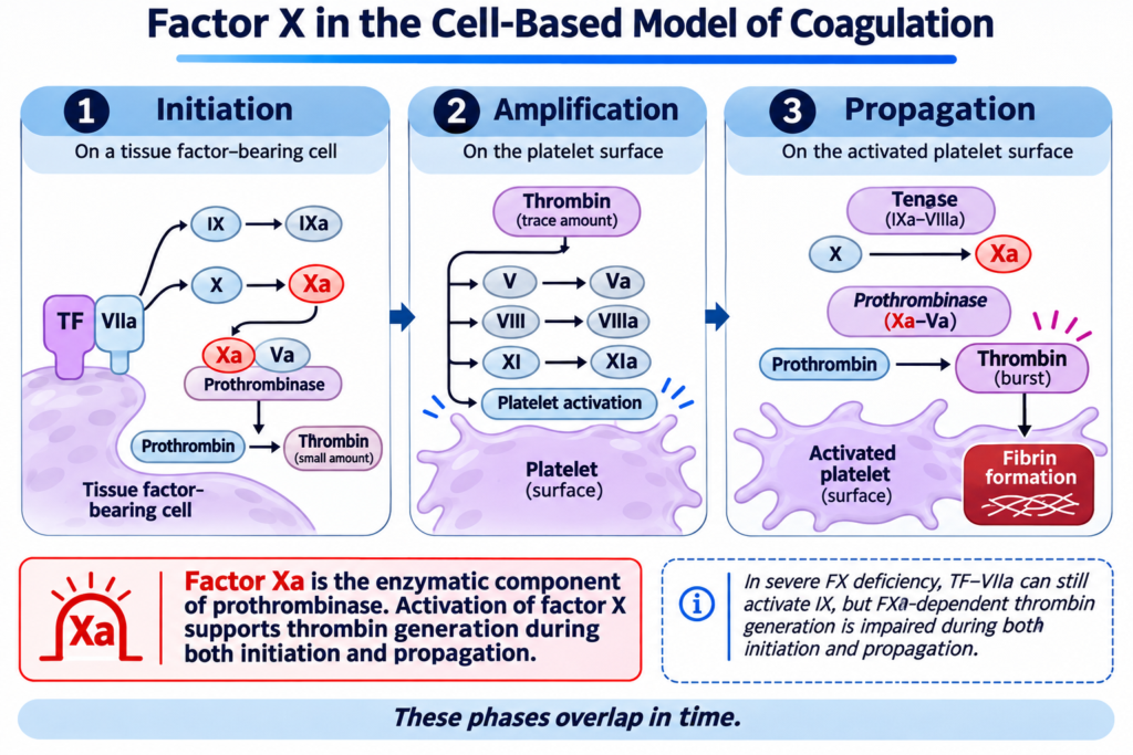
This spectrum reflects the position of factor X within the coagulation system. Factor X is activated by both the tissue factor-dependent pathway and the intrinsic tenase complex. Once activated, it combines with factor Va to form prothrombinase, which generates thrombin. Unlike a defect confined predominantly to one route of activation, severe FX deficiency compromises a critical component shared by both.
The relationship between FX activity and bleeding severity is therefore particularly instructive. Among rare inherited coagulation deficiencies, FX activity is one of the factor measurements most closely associated with the clinical phenotype. Nevertheless, the association is not absolute. Some patients bleed more than their measured activity might suggest, while others experience fewer complications than expected.
Factor X deficiency also has an important acquired counterpart. In systemic immunoglobulin light-chain (AL) amyloidosis, circulating FX may be removed from plasma through interactions with amyloid deposits. The resulting laboratory abnormality can resemble congenital deficiency, even though the underlying mechanism and clinical context are entirely different.
Understanding factor X deficiency requires connecting three questions: Why does this particular deficiency cause severe bleeding? How much does residual activity tell us about an individual patient? And what mechanism explains the low activity?
1. Why does factor X occupy such an important position in hemostasis?
Factor X is a vitamin K-dependent plasma glycoprotein synthesized primarily by hepatocytes. It circulates as an inactive precursor, or zymogen, and must undergo proteolytic activation before it can participate directly in thrombin generation.
Its importance lies in the convergence of two major routes of coagulation activation.
Following vascular injury, exposure of tissue factor allows the tissue factor-FVIIa complex to activate FX. This initiates thrombin generation and helps establish the early hemostatic response.
As coagulation proceeds, factor IXa combines with factor VIIIa on a phospholipid membrane to form the intrinsic tenase complex. This complex also activates FX, supporting the amplification and propagation of thrombin generation.
Both reactions produce activated factor X (FXa). In the presence of factor Va, calcium, and an appropriate phospholipid surface, FXa forms the prothrombinase complex. Prothrombinase converts prothrombin to thrombin much more efficiently than FXa acting alone.
Thrombin then performs several essential tasks: it converts fibrinogen to fibrin, activates platelets, and activates additional coagulation factors that support a sustained hemostatic response.
This creates a fundamental difference between factor X and a factor involved principally in an upstream pathway.
When factor X is severely deficient, both major routes of activation may remain intact upstream, but their ability to generate thrombin is constrained by insufficient FX substrate.
The consequences are amplified by the catalytic role of prothrombinase. Factor Xa is not simply another component in a linear series of reactions. Its association with factor Va creates a highly efficient enzyme complex capable of generating large quantities of thrombin at the site of injury.
Factor X deficiency can therefore impair the formation of the initial thrombin signal and the subsequent thrombin burst. The resulting bleeding phenotype reflects compromised initiation and propagation rather than a defect isolated to one conventional laboratory pathway.12
2. How is factor X constructed, and what can go wrong?
Factor X is encoded by F10, located on chromosome 13q34 near F7, the gene encoding factor VII. Congenital FX deficiency generally follows autosomal recessive inheritance and affects males and females.
The estimated prevalence of severe inherited FX deficiency has traditionally been approximately one individual per million. However, this estimate is uncertain and probably underestimates milder or asymptomatic biochemical deficiency. The disorder is more frequently recognized in populations where consanguinity increases the likelihood of inheriting the same rare pathogenic variant from both parents.34
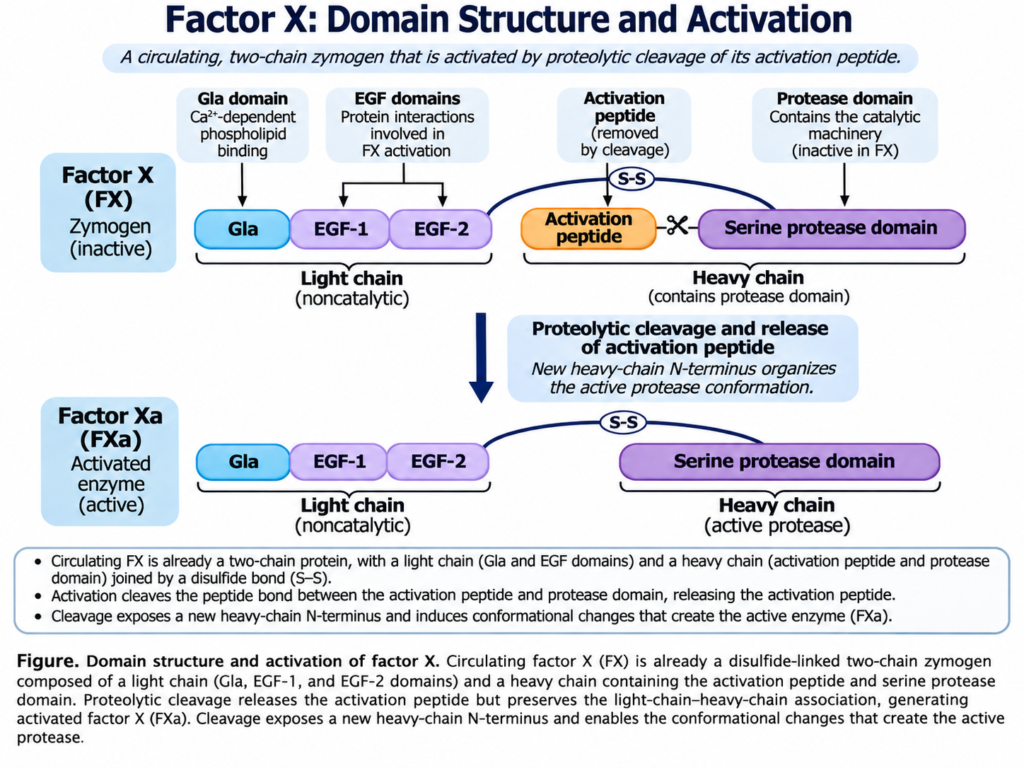
The mature FX molecule contains two polypeptide chains connected by a disulfide bond. The light chain contains a gamma-carboxyglutamic acid (Gla) domain and two epidermal growth factor-like domains. The heavy chain contains the activation peptide and catalytic serine protease domain.
Each region contributes to the protein’s function.
- Gla domain: Vitamin K-dependent gamma-carboxylation allows calcium-dependent interactions with negatively charged phospholipid membranes.
- Epidermal growth factor-like domains: These contribute to the molecule’s structure and interactions with other components of coagulation.
- Activation peptide: Proteolytic cleavage permits conversion of the circulating zymogen into FXa.
- Catalytic domain: This supports the enzymatic activity of FXa, including conversion of prothrombin when FXa is assembled into prothrombinase.
Before secretion, FX also undergoes intracellular processing, including removal of signal sequences, posttranslational modification, and formation of its mature two-chain structure.
Pathogenic variants can interfere with these processes at several points. Some reduce protein synthesis or prevent normal secretion. Others produce circulating FX that is structurally abnormal, cannot be activated efficiently, interacts poorly with its cofactors or substrates, or has impaired catalytic activity.
The resulting defects can be described as quantitative or qualitative.
| Type of deficiency | FX coagulant activity (FX) | FX antigen (FX) | Principal biological interpretation |
|---|---|---|---|
| Type I, quantitative | Reduced | Reduced, generally to a similar extent | Too little FX protein reaches the circulation |
| Type II, qualitative | Reduced | Relatively preserved | Circulating FX is present but functions abnormally |
| Mixed quantitative and qualitative | Reduced | Reduced to a lesser or variable extent | Both protein abundance and intrinsic function contribute |
This classification describes the protein defect, not the patient’s clinical bleeding severity. A patient with type II deficiency may have a relatively preserved antigen concentration but severely impaired coagulant activity. In type I deficiency, reduced protein abundance leads to reduced coagulant activity, but the resulting bleeding phenotype is not determined by factor activity alone.
The distinction also matters diagnostically. A low FX activity identifies a functional abnormality, but the antigen result helps establish whether the underlying defect involves protein quantity, function, or both.5
3. What do F10 variants reveal about the relationship between molecular structure and disease?
Most reported pathogenic F10 variants are missense variants, although nonsense, frameshift, splice-site, and larger deletion variants have also been described. Many are family-specific, making it difficult to associate an individual variant with a reproducible clinical outcome.
Nevertheless, molecular studies reveal several distinct mechanisms of disease.
Variants affecting the signal peptide may interfere with normal intracellular processing and secretion. Variants in the Gla domain may impair calcium-dependent membrane interactions. Others alter structural domains or the catalytic site, producing circulating protein with reduced function.
Pastoret and colleagues investigated 16 families with hereditary FX deficiency using high-throughput sequencing. They identified 19 sequence variants and two copy-number abnormalities, including five previously unreported variants.6
Several of these findings are particularly informative.
One variant, p.Glu22Val, was predicted to interfere with cleavage of the signal peptide, potentially reducing secretion of mature FX. Another, p.Cys342Tyr, was predicted to disrupt the disulfide bond connecting the heavy and light chains. A third, p.Val385Met, affected the catalytic protease domain.
These variants illustrate how different molecular abnormalities can converge on reduced FX activity. Importantly, the proposed mechanisms for some newly identified variants were based partly on structural modeling and computational prediction, rather than direct functional demonstration.
The same study also identified two individuals with combined FVII and FX deficiency involving chromosome 13q34 deletions. In these patients, loss of one copy of the region containing F7 and F10 occurred together with a variant affecting the remaining F10 allele.
This explains why FX activity could be substantially lower than FVII activity. The deletion removed one copy of each gene, but the remaining F7 allele was intact, whereas the remaining F10 allele carried an additional pathogenic variant. Thus, FVII activity was only partially reduced, while FX function was impaired by abnormalities affecting both gene copies.
Combined FVII/FX deficiency should therefore not automatically be interpreted as two unrelated inherited disorders. The close physical location of F7 and F10 creates the possibility of a shared structural genetic abnormality.7
Genotype helps explain the biological origin of deficiency, but it does not eliminate variability in bleeding.
A patient with variants on both F10 alleles generally has lower activity and greater bleeding risk than a heterozygous relative. Yet the presence of a particular variant does not establish exactly when, where, or how severely an individual will bleed.
That distinction becomes clearer when factor activity is considered alongside the clinical phenotype.
4. What is the clinical bleeding phenotype of congenital factor X deficiency?
Congenital FX deficiency spans a broad clinical spectrum, but its severe forms are among the most consequential of the rare inherited coagulation disorders.
Patients with profound deficiency may present during the neonatal period or early infancy. Umbilical stump bleeding is a characteristic presentation, sometimes accompanied by gastrointestinal hemorrhage, intracranial bleeding, or bleeding after minor procedures.
These events can occur before the child has experienced the usual hemostatic challenges of later childhood.
The timing is important. Normal newborns have developmentally lower concentrations of several vitamin K-dependent coagulation factors, including FX, than healthy adults. A child with profound inherited deficiency begins life with an already severely limited capacity to generate thrombin. The physiological characteristics of neonatal hemostasis and the demands imposed by birth can make this a particularly vulnerable period.
Because FX concentrations are physiologically lower at birth than in adulthood, laboratory interpretation in a newborn requires age-appropriate reference intervals. A value below the adult reference range does not, by itself, establish congenital FX deficiency. Profoundly reduced activity in an infant with significant bleeding nevertheless warrants urgent investigation, particularly when the abnormality is persistent or supported by family history.8
The manifestations of congenital FX deficiency include:
- Neonatal and early-life bleeding: Umbilical stump hemorrhage, intracranial hemorrhage, gastrointestinal bleeding, and excessive bleeding following procedures.
- Musculoskeletal bleeding: Hemarthroses and muscle hematomas, sometimes producing chronic joint damage or functional impairment.
- Mucocutaneous bleeding: Epistaxis, gum bleeding, bruising, and bleeding after dental extraction.
- Gastrointestinal and genitourinary bleeding: Hemorrhage involving the gastrointestinal tract or urinary system, particularly in more severe disease.
- Reproductive bleeding: Heavy menstrual bleeding, ovulation-associated hemorrhage, and postpartum bleeding.
- Procedure-related bleeding: Excessive bleeding after surgery, trauma, or other hemostatic challenges, including in some individuals with less severe baseline deficiency.
A 2021 review of severe bleeding in hereditary FX deficiency identified intracranial hemorrhage as the most frequently reported severe manifestation in the selected literature. Among 197 patients in publications reporting specific bleeding sites, 82 had central nervous system bleeding, 51 had hemarthrosis, 31 had gastrointestinal bleeding, and 23 had umbilical bleeding.9
These figures are useful for understanding the spectrum of published severe cases, but they must not be interpreted as population prevalences. The review deliberately selected publications describing severe bleeding, creating substantial ascertainment and publication bias.
The clinical importance of neonatal intracranial hemorrhage cannot be overstated. It may be the first recognized manifestation of the disorder, and surviving children may experience permanent neurological consequences.
A separate review identified 45 reported patients with FX deficiency and intracranial hemorrhage, with 30 events occurring before 19 months of age. Although the cases were highly selected, they reinforce the particular vulnerability of infancy.10
The phenotype is not restricted to infancy. Adults with severe deficiency may experience repeated joint or muscle bleeding, while those with intermediate activity may first become symptomatic after surgery, trauma, or childbirth.
Conversely, some patients with mild biochemical deficiency have little spontaneous bleeding. Their diagnosis may follow a family investigation or routine coagulation testing rather than a major hemorrhagic event.
The bleeding history must therefore establish not only whether bleeding has occurred but also the patient’s opportunities to demonstrate hemostatic competence.
5. Why is factor X activity more closely associated with bleeding severity than many other rare factor deficiencies?
The relationship between residual factor activity and clinical bleeding differs substantially among rare coagulation disorders.
In FVII or FXI deficiency, patients with very low activity may experience surprisingly little bleeding, and factor activity alone has limited precision in predicting the individual phenotype.
Factor X deficiency behaves differently.
In the European Network of Rare Bleeding Disorders (EN-RBD) study, factor X was among the coagulation factors whose activity showed the strongest relationship with clinical bleeding severity, alongside fibrinogen and FXIII.11
The study included 489 patients with rare inherited bleeding disorders, of whom 45 had FX deficiency. Bleeding phenotypes were categorized as asymptomatic, bleeding after hemostatic challenges, minor spontaneous bleeding, or major spontaneous bleeding.
Lower FX activity was associated with more severe clinical bleeding in the EN-RBD study, with activity below approximately 10 IU/dL associated with major spontaneous hemorrhage. The analysis also estimated an FX activity of approximately 56 IU/dL in association with the asymptomatic category.12 A subsequent ISTH SSC classification proposed FX activity above 40 IU/dL for the mild, largely asymptomatic category.13 These are different kinds of numbers: the first is a study-derived association, whereas the second is an operational classification boundary. Neither should be interpreted as a precise predictor of bleeding in an individual patient.
This finding is biologically plausible. Since both major routes of coagulation activation require FX, low residual activity constrains thrombin generation regardless of which pathway initiates the response. At higher residual activity, some patients may generate sufficient thrombin for ordinary hemostatic challenges even though laboratory activity remains below the population reference range.
However, the activity-severity relationship should not be interpreted as a precise physiological boundary.
Historical classifications of FX deficiency have used thresholds derived largely from hemophilia, but their definitions have not been uniform.
| Framework | Severe | Moderate | Mild |
|---|---|---|---|
| Selected historical FX laboratory classifications | Below 1 IU/dL | 1 to 5 IU/dL | Approximately 6 to 10 IU/dL in some schemes |
| EN-RBD phenotype-informed classification | Below 10 IU/dL | 10 to 40 IU/dL | Above 40 IU/dL |
Historical FX studies did not use a uniform mild-deficiency boundary; some classified all activity above 5 IU/dL as mild. The EN-RBD categories instead reflect associations with observed bleeding severity. Neither system is a validated individual prediction rule.
The difference between the schemes is substantial. Under a historical scheme using 6 to 10 IU/dL as mild deficiency, an activity of 7 IU/dL would be described as mild. Under the phenotype-informed scheme, it lies in a range associated with increased risk of major spontaneous bleeding.
The classification is therefore not merely a matter of terminology. It determines what the label severe is intended to communicate.
The EN-RBD approach attempts to connect the laboratory measurement with clinical risk rather than borrowing a classification developed for different disorders.14
There are limitations. The number of FX-deficient patients in the original EN-RBD analysis was relatively small. Patients were recruited through specialized centers, and the classification was derived from observed phenotypes rather than prospectively tested against future bleeding.
Moreover, the most severely affected individuals may have received replacement or prophylaxis, potentially altering the subsequent bleeding history.
The principal conclusion is therefore probabilistic:
As FX activity falls, especially below approximately 10 IU/dL, the likelihood of serious bleeding increases. But the activity does not independently establish the outcome of a particular future challenge.
This is a stronger association than is observed in some other rare factor deficiencies, but it remains an association between a laboratory measurement and a clinical phenotype, not a deterministic law.
Evidence Box: What Does Factor X Activity Tell Us About Bleeding Severity?
| Evidence | Principal finding | Interpretation and limitation |
|---|---|---|
| EN-RBD study, 2012 | FX activity showed one of the strongest associations with bleeding severity among the rare factor deficiencies studied. | Supports activity-informed phenotype classification; based on relatively few FX-deficient patients. |
| Karimi et al., 2008 | Ten homozygous patients with severe FX deficiency had substantial bleeding morbidity, while some heterozygous relatives also reported bleeding after procedures or delivery. | Illustrates both severe disease and exceptions among carriers; small, highly selected family cohort. |
| Tarantino, 2021 | Published severe bleeding cases frequently involved intracranial, joint, gastrointestinal, or umbilical bleeding, often in early childhood. | Documents the spectrum of severe disease; not suitable for estimating population incidence of specific manifestations. |
| Pastoret et al., 2023 | Patients with biallelic F10 abnormalities generally had lower activity and more severe bleeding than most heterozygous relatives. | Supports a genotype-activity-phenotype relationship, but molecular findings do not determine individual severity. |
The evidence supports a clinically meaningful association between FX activity and bleeding severity while preserving uncertainty in individual risk prediction.
6. What special reproductive bleeding patterns occur in women with factor X deficiency?
Factor X deficiency affects males and females, but women encounter additional hemostatic challenges through menstruation, ovulation, pregnancy, and childbirth.
Heavy menstrual bleeding is common in affected women, including some with relatively mild biochemical deficiency. Its consequences may include iron deficiency, anemia, and repeated need for medical care.
Ovulation-associated hemorrhage is particularly important because bleeding into a corpus luteum cyst or the peritoneal cavity may produce substantial blood loss. This complication can be recurrent and may occur independently of pregnancy or childbirth.
Pregnancy also raises distinct concerns. Although coagulation factor concentrations change physiologically during pregnancy and FX activity may increase, the rise may not be sufficient to correct a severe inherited defect. Postpartum hemorrhage remains an important complication.
In a systematic review of congenital FX deficiency in women, Spiliopoulos and Kadir identified 49 publications describing 332 women. Heavy menstrual bleeding was reported in 72 of 284 women with relevant data, or 25%. Ovulation-related hemoperitoneum requiring transfusion was reported in eight of 322 women, and postpartum hemorrhage occurred in six of 27 reported deliveries.15
These denominators reflect different subsets of the literature. The review combined case reports and small series with heterogeneous definitions and ascertainment. Consequently, the percentages should be interpreted as frequencies among reported patients, not as reliable estimates for all women with congenital FX deficiency.
An additional consideration is that women carrying a single pathogenic F10 variant may also report excessive bleeding after delivery or other hemostatic challenges. Such observations do not establish that every heterozygous carrier has a clinically important bleeding disorder, but they caution against assuming that heterozygosity is necessarily irrelevant.
The reproductive phenotype therefore reinforces the central lesson of congenital FX deficiency: residual activity helps characterize bleeding risk, while the type and extent of hemostatic exposure influence how that risk becomes clinically apparent.
Specific approaches to menstrual bleeding, pregnancy, delivery, and prophylactic replacement belong in the companion treatment essay.
7. How is factor X deficiency identified in the coagulation laboratory?
Because FX participates in the common pathway of coagulation, congenital deficiency typically prolongs both the prothrombin time (PT) and the activated partial thromboplastin time (aPTT).
The PT initiates coagulation through the tissue factor-dependent pathway. The aPTT uses a different activation system that engages the intrinsic pathway. Both assays ultimately require FX activation and the generation of thrombin.
Severe FX deficiency can therefore impair both tests, although the degree of prolongation depends on residual activity and reagent sensitivity.
This pattern is not specific for FX deficiency. Deficiencies of other common-pathway factors, combined coagulation factor abnormalities, anticoagulants, and several acquired disorders may produce similar results.
The characteristic evaluation includes a specific measurement of FX coagulant activity (FX), usually using a one-stage clotting assay with FX-deficient substrate plasma.
Such assays may use PT-based or aPTT-based methods. The observed clotting time is compared with calibrated reference plasma to estimate the activity contributed by the patient’s FX.
Additional testing can help characterize unusual findings.
FX antigen (FX) measures circulating protein and helps distinguish quantitative from qualitative deficiency.
Chromogenic FX assays measure FX function through an enzyme-based system, generally involving FX activation followed by measurement of FXa activity against a chromogenic substrate.
Russell’s viper venom-based assays exploit the ability of venom-derived enzymes to activate FX through a mechanism different from physiological tissue factor-dependent or intrinsic tenase-dependent activation.
These methods can help determine whether the abnormality reflects impaired FX quantity, activation, or catalytic function.1617
An important analytical limitation is interference from anticoagulants, particularly direct factor Xa inhibitors such as apixaban and rivaroxaban. These drugs can produce falsely reduced FX activity in one-stage clotting assays. They may also interfere with chromogenic FX assays because the measured reaction depends on FXa activity. Changing from a clot-based to a chromogenic assay does not necessarily eliminate direct FXa inhibitor interference. Medication exposure, specimen timing, and assay-specific susceptibility must therefore be considered before concluding that reduced measured FX activity represents a true endogenous deficiency. When necessary, specialist laboratory methods can help establish whether an anticoagulant is responsible.18
A particularly important limitation is that different activity assays do not interrogate precisely the same molecular reaction.
A qualitative FX variant may interact abnormally with one physiological activation complex while retaining function in another experimental system. Consequently, PT-based, aPTT-based, and venom-based FX assays may produce discrepant activity results.
This is not necessarily a laboratory error. It may reflect the particular reaction that the variant impairs.
Factor X Padua provides a useful example. This qualitative variant predominantly impairs FX activation through the tissue factor-dependent pathway. A PT-based functional assay may therefore yield substantially reduced activity, while assays using other routes of FX activation produce different results. The molecular defect involves substitution of tryptophan for arginine at position 251 of mature FX, also designated Arg291Trp when the full precursor is numbered. Molecular modeling suggests that the substitution affects a calcium-binding region without substantially destabilizing the protein, although this proposed structural mechanism should not be confused with direct experimental proof of every functional consequence.1920
The phenotype may therefore be described more precisely when the assay result is linked to the mechanism being measured.
For example, a variant that impairs activation by tissue factor-FVIIa may produce a different functional result from one that disrupts the catalytic activity of FXa itself. The latter defect would be expected to affect thrombin generation more broadly.
The interpretation of assay discrepancies belongs within the biological characterization of FX deficiency, rather than treating every abnormal measurement as an interchangeable estimate of a single underlying quantity.
8. How should qualitative variants and genetic testing be interpreted?
Molecular testing may identify the cause of persistent congenital FX deficiency, support family investigation, and clarify unusual laboratory findings. It can also distinguish a single F10 sequence variant from a larger chromosome 13q34 deletion involving both F10 and F7.
The diagnostic value of sequencing depends on the question being asked.
In a child with profound FX deficiency, severe spontaneous bleeding, and an affected sibling, identifying biallelic pathogenic F10 variants can establish the molecular explanation and support genetic counseling.
In an adult with modestly reduced activity, little bleeding, and a single heterozygous variant, the result may be less definitive. The variant’s functional consequences, the patient’s bleeding history, and the presence of additional abnormalities remain relevant.
The Pastoret study demonstrates several benefits of broader molecular investigation. High-throughput sequencing identified sequence variants and copy-number abnormalities, including structural deletions that would not necessarily be recognized by conventional sequence-only testing.
It also identified variants associated with qualitative abnormalities, in which FX antigen and functional activity were not reduced to the same degree.21
Several cautions follow.
First, a predicted molecular mechanism is not always experimentally established. Structural modeling may support pathogenicity, but direct functional studies provide different evidence.
Second, finding a variant does not establish that it explains every bleeding manifestation. Another bleeding disorder, a concurrent acquired abnormality, or an unrelated cause of bleeding may coexist.
Third, a negative sequencing result does not exclude every inherited mechanism. Copy-number abnormalities, difficult-to-detect structural variants, or regulatory variants may require additional testing.
Finally, not every patient with low FX activity needs comprehensive molecular characterization before a clinical diagnosis can be made. Persistent selective deficiency, clinical findings, and family evidence may be sufficient to establish a working diagnosis, even while molecular confirmation remains incomplete.
Genetic testing explains the origin of a deficiency most effectively when its findings are interpreted together with the biochemical phenotype and the clinical history.
9. How does acquired factor X deficiency differ from congenital disease?
Factor X deficiency may also develop in individuals who previously had normal hemostasis. Acquired deficiency is mechanistically diverse and should not be treated as a single disorder.
True acquired reductions in FX activity may accompany vitamin K deficiency, vitamin K antagonist therapy, liver disease, and consumptive coagulopathy. In these settings, FX is usually one of several coagulation proteins affected. Direct factor Xa inhibitors introduce a different problem: they can produce apparently low FX activity through assay interference without reducing the amount of endogenous FX protein.
The more distinctive diagnostic problem is isolated acquired factor X deficiency, in which FX activity is disproportionately reduced relative to other factors.
Acquired Factor X Deficiency in AL Amyloidosis
The best-recognized association is systemic AL amyloidosis.
AL amyloidosis arises from a clonal plasma cell disorder that produces immunoglobulin light chains capable of forming amyloid fibrils. These deposits accumulate in tissues and may cause organ dysfunction.
Acquired FX deficiency in AL amyloidosis has traditionally been attributed to adsorption of circulating FX onto amyloid fibrils, accelerating its removal from plasma. Additional mechanisms involving FX interactions with amyloid-associated proteins and macrophage clearance have been investigated.
The essential distinction is that the patient’s F10 gene may be normal and hepatic synthesis may continue, yet circulating FX activity falls because removal from plasma is increased.
Thus, the same low FX activity can result from inadequate production of functional protein in congenital deficiency or accelerated clearance of otherwise functional protein in acquired disease.22
Acquired FX deficiency is not present in every patient with AL amyloidosis. Earlier reports suggested that approximately 6% to 14% of affected patients had reduced FX activity, although the estimates depended on the laboratory cutoff used and the populations studied.
A contemporary retrospective study from the Boston University Amyloidosis Center evaluated 1,956 patients with AL amyloidosis from 2000 through 2023. Eighty-eight, or approximately 4.5%, had FX activity of 50% or less after exclusion of patients receiving anticoagulant therapy.
Among the 88 patients with acquired FX deficiency, 49 (56%) had at least one bleeding manifestation at or near diagnosis. Bleeding was reported in 44% of those with FX activity of 41% to 50%, 64% with activity of 21% to 40%, and 71% with activity of 20% or less.
Although bleeding tended to be more frequent with lower activity, the differences were not statistically significant in this cohort.
Longitudinal FX measurements were available for a selected subset of 28 of the 88 patients with acquired deficiency. Within this subset, patients who achieved complete hematologic response had greater improvements in FX activity than those who did not. The limited follow-up population introduces potential selection and surveillance bias, and FX activity also normalized in two patients without a complete hematologic response. These observations support a relationship between control of the underlying plasma cell disorder and FX recovery without establishing that clonal response is its sole determinant.23
This observation supports the concept that controlling the amyloidogenic plasma cell clone can affect the underlying mechanism of FX loss. The treatment implications belong in the relevant disease-management context.
It is also important not to attribute all bleeding in AL amyloidosis to FX deficiency. Amyloid-associated vascular fragility, tissue infiltration, hepatic involvement, and other hemostatic abnormalities may contribute to bleeding independently of the measured FX activity.
A low FX measurement in a patient with AL amyloidosis is therefore clinically meaningful but not necessarily the sole explanation for the observed bleeding.
Acquired factor X inhibitors
A much less common acquired mechanism involves antibodies directed against FX.
These antibodies may interfere directly with coagulant function or promote accelerated clearance. The resulting presentation can include sudden, severe bleeding with no previous personal or family history of abnormal hemostasis.
Ichinose and colleagues identified 54 reported patients with suspected autoimmune FX deficiency, classifying eight as definite, 18 as probable, and 28 as possible cases. Their detailed analysis focused on the 26 definite or probable cases, distinguished from amyloid-associated deficiency and other acquired causes.24
An important finding was that some affected patients had low or absent measurable neutralizing inhibitor activity despite evidence of anti-FX autoantibodies. This suggests that antibody-mediated clearance, rather than direct inhibition in a conventional clotting assay, may contribute in selected cases.
A mixing study primarily examines whether the patient’s plasma inhibits coagulation under the conditions of an in vitro assay. An antibody that accelerates FX clearance in vivo may not produce a conventional inhibitory pattern. Consequently, correction with normal plasma does not invariably exclude antibody-associated acquired FX deficiency. When the clinical findings remain suspicious, specialist assessment may require direct investigation for anti-FX antibodies as well as functional inhibitor testing.25
Nevertheless, acquired FX inhibitors remain exceptionally rare. The clinical context, persistence of the abnormality, and results of targeted testing are essential before attributing low FX activity to an autoimmune process.
10. How should congenital and acquired factor X deficiencies be distinguished?
The diagnosis begins with the laboratory finding but requires an explanation for why FX activity is reduced.
A persistent, selective deficiency in a child with early-life bleeding and an affected sibling suggests a congenital disorder. An older patient with a newly prolonged PT and aPTT, previously normal coagulation studies, and evidence of a plasma cell disorder presents a different problem.
The laboratory phenotype may overlap, but the etiologic evidence differs.
| Finding | Congenital FX deficiency | AL amyloidosis-associated acquired FX deficiency | Autoimmune acquired FX deficiency |
|---|---|---|---|
| Typical context | Early-life bleeding, family history, or incidental testing | Established or newly recognized AL amyloidosis | New bleeding without a congenital history |
| Primary mechanism | Pathogenic F10 variant affecting quantity or function | Accelerated loss of circulating FX associated with amyloid deposits | Antibody-related impairment of FX function or clearance |
| PT and aPTT | Often both prolonged, depending on activity | May both be prolonged | May both be prolonged |
| FX activity | Reduced | Reduced | Reduced |
| FX antigen | Reduced or relatively preserved, depending on variant | Often reduced because circulating protein is lost | Variable, depending on antibody mechanism |
| Genetic findings | Pathogenic F10 variants may establish molecular diagnosis | No F10 pathogenic variant required | No F10 pathogenic variant required |
| Additional evidence | Family testing, persistent deficiency, molecular studies | Evidence of a plasma cell clone and amyloid deposition | Inhibitor or anti-FX antibody studies, interpreted in context |
The table illustrates why diagnosing a low FX activity requires more than recognizing a common-pathway screening pattern.
Before attributing a reduced activity to any of these mechanisms, the clinician must also establish that the result reflects genuine endogenous FX deficiency rather than anticoagulant-related assay interference or another analytical limitation.
For congenital deficiency, the investigation may emphasize molecular findings, family studies, and the relationship between FX activity and phenotype.
For amyloid-associated deficiency, the more important clue may be an acquired abnormality in a patient with findings suggesting systemic amyloidosis.
For autoimmune deficiency, the evaluation may require specialized testing because ordinary inhibitor assays do not identify every antibody-mediated mechanism.
As in other rare factor deficiencies, the final diagnosis should distinguish three questions: whether the abnormality is real, what has caused it, and how much it contributes to the patient’s bleeding.
The answer to the third question may remain uncertain, especially when other acquired hemostatic abnormalities coexist.
Reflect and Apply
Consider two patients with similarly reduced FX activity.
Patient A is a 3-month-old infant referred after excessive umbilical stump bleeding and an intracranial hemorrhage. PT and aPTT are prolonged, and FX activity is 3 IU/dL. Other coagulation factor activities are not comparably reduced. The parents report that an older sibling experienced severe bleeding during infancy.
Patient B is a 66-year-old adult with easy bruising, fatigue, and newly abnormal coagulation studies. PT and aPTT are prolonged, and FX activity is 12 IU/dL. Coagulation testing performed several years earlier was normal. Further evaluation identifies a monoclonal lambda light chain and biopsy-confirmed systemic AL amyloidosis.
Consider the following questions:
- What does the shared PT/aPTT pattern establish, and what does it fail to distinguish?
- How does the position of FX within coagulation help explain Patient A’s severe early-life bleeding?
- How should the measured FX activity influence interpretation of bleeding severity in each patient?
- What mechanisms could account for reduced FX activity in Patient A and Patient B?
- What additional laboratory or molecular findings would strengthen the causal explanation in each case?
- Why might Patient B’s bleeding phenotype not be fully explained by the FX activity alone?
Closing Thought
Factor X deficiency demonstrates how the biological position of a coagulation protein shapes the consequences of its loss. Factor X is activated by two major pathways but becomes the common enzymatic link to thrombin generation. Profound deficiency can therefore compromise hemostasis from the earliest stages of life, and residual FX activity provides meaningful information about bleeding severity.
Yet the laboratory value is not a complete clinical explanation. Quantitative and qualitative variants may produce similar activity measurements through different mechanisms, while inherited and acquired deficiencies may produce nearly identical screening patterns. In AL amyloidosis, the problem may be accelerated disappearance of a normal protein rather than failure to produce it.
Factor X activity describes an important functional limitation. The molecular or acquired mechanism explains why the limitation exists. The clinical phenotype reveals its consequences. A diagnosis becomes meaningful when all three are understood together.