
Copper deficiency can cause anemia.
At first, that may seem surprising. Copper is not incorporated into hemoglobin, and unlike iron, vitamin B12, or folate, its connection to red-cell production is not immediately obvious.
The explanation is not a single pathway. Copper participates in iron handling, mitochondrial respiration, antioxidant defense, heme synthesis, and hematopoietic cell development. When copper becomes deficient, several of these processes may be disturbed at once.1
That helps explain the unusually broad phenotype. Copper-deficiency anemia may be microcytic, normocytic, or macrocytic. Neutropenia is common. Thrombocytopenia can occur. The marrow may show vacuolated precursors, increased iron, or ring sideroblasts, and the overall picture can resemble myelodysplastic syndrome.
Understanding the syndrome begins with what copper normally does.
What does copper normally do?
Copper is an essential trace element that serves as a cofactor for enzymes involved in several fundamental cellular processes. Four functions are particularly relevant to hematopoiesis.
Iron handling. Copper is required for the multicopper ferroxidases ceruloplasmin and hephaestin. Both catalyze oxidation of Fe²⁺ to Fe³⁺, helping iron exported through ferroportin enter the transferrin-bound plasma pool.2
Mitochondrial respiration. Copper is an essential component of cytochrome c oxidase (complex IV), the terminal enzyme of the mitochondrial electron-transport chain. Copper is therefore required for normal oxidative phosphorylation and mitochondrial energy production.3
Antioxidant defense. Copper is a cofactor for Cu/Zn superoxide dismutase (SOD1), which converts superoxide into less reactive products and helps protect cells from oxidative injury.4
Hematopoietic cell development. Experimental studies suggest that copper availability influences the differentiation and survival of hematopoietic progenitors, including erythroid and myeloid cells.5
Copper also participates in processes outside hematopoiesis, including connective-tissue formation and nervous-system function. Those roles help explain some of the nonhematologic manifestations of copper deficiency.
Key copper-dependent proteins relevant to hematopoiesis:
| Protein | Copper-dependent function | Hematologic relevance |
|---|---|---|
| Ceruloplasmin | Multicopper ferroxidase | Helps mobilize recycled and stored iron into the transferrin pool |
| Hephaestin | Membrane-associated multicopper ferroxidase | Facilitates iron export from intestinal enterocytes |
| Cytochrome c oxidase (complex IV) | Electron transfer and oxidative phosphorylation | Supports mitochondrial function in developing blood cells |
| Cu/Zn superoxide dismutase (SOD1) | Antioxidant defense | Limits oxidative cellular injury |
The first two proteins provide a particularly direct connection between copper and erythropoiesis: copper helps determine whether iron can move efficiently from cells into plasma.
Why does copper deficiency cause anemia?
There is probably no single mechanism.
Each of the normal copper-dependent functions described above provides a potential route from copper deficiency to impaired erythropoiesis. The best characterized involves iron trafficking, but mitochondrial dysfunction, altered heme synthesis, oxidative stress, and effects on hematopoietic progenitors may also contribute.6
Iron handling
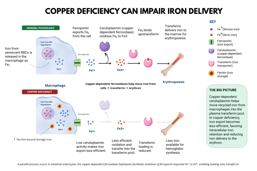
Copper-dependent ferroxidases help iron make the transition from inside cells into the transferrin-bound plasma pool that supplies the marrow.
Iron leaves cells through ferroportin as ferrous iron, Fe²⁺.
Transferrin binds ferric iron, Fe³⁺.
The exported iron therefore undergoes oxidation:
Fe²⁺ → Fe³⁺
The copper-dependent ferroxidases hephaestin and ceruloplasmin catalyze this reaction.7
HOW DOES COPPER ALLOW CERULOPLASMIN TO OXIDIZE IRON?
Ceruloplasmin is a multicopper oxidase enzyme. Copper atoms are incorporated into specific catalytic sites within the protein and shuttle electrons during the ferroxidase reaction.
Fe²⁺ → Fe³⁺ + e⁻
The electron is transiently accepted by a copper center:
Cu²⁺ + e⁻ → Cu⁺
The electrons are ultimately transferred to molecular oxygen:
O₂ + 4H⁺ + 4e⁻ → 2H₂O
Overall:
4Fe²⁺ + O₂ + 4H⁺ → 4Fe³⁺ + 2H₂O
The copper atoms therefore act as catalytic electron-transfer centers. They cycle between oxidation states but are not consumed in the reaction.
But the oxidation-state change is not merely an interesting piece of chemistry. It helps couple cellular iron export to plasma iron transport.
Normal iron export
Intracellular Fe²⁺ → ferroportin → extracellular Fe²⁺ → ferroxidase → Fe³⁺ → transferrin
When copper-dependent ferroxidase activity is reduced, iron export and mobilization become less efficient, favoring intracellular iron retention and reducing transfer of iron into the circulating transferrin pool.8
Copper deficiency
↓ copper → ↓ ferroxidase activity → ↓ efficient iron export/mobilization → ↑ intracellular iron retention → ↓ iron entering transferrin pool
The same chemistry operates in two important physiologic settings.
Dietary iron
Enterocyte Fe²⁺ → ferroportin → Fe²⁺ → hephaestin → Fe³⁺ → transferrin
Hephaestin is a membrane-associated multicopper ferroxidase concentrated near the basolateral surface of intestinal enterocytes. Its position places ferroxidase activity directly at the site where newly absorbed dietary iron leaves the cell.9
Recycled iron
Macrophage Fe²⁺ → ferroportin → Fe²⁺ → ceruloplasmin → Fe³⁺ → transferrin
Ceruloplasmin is predominantly a soluble circulating multicopper ferroxidase and is particularly important in mobilizing recycled and stored iron from cells such as macrophages.10
Hephaestin and ceruloplasmin: same chemistry, different emphasis:
| Hephaestin | Ceruloplasmin | |
|---|---|---|
| Protein type | Multicopper ferroxidase | Multicopper ferroxidase |
| Major form | Membrane-associated | Mainly soluble circulating; membrane-associated forms also exist |
| Major physiologic setting | Intestinal iron export | Systemic iron mobilization |
| Particularly important at | Basolateral enterocyte | Macrophages and other iron-exporting tissues |
| Reaction | Fe²⁺ → Fe³⁺ | Fe²⁺ → Fe³⁺ |
| Relationship to ferroportin | Positioned locally at the iron-export site | Oxidizes ferroportin-exported iron extracellularly |
| Copper dependent | Yes | Yes |
Hephaestin and ceruloplasmin should not be viewed as absolutely compartment-specific. Their functions overlap, but hephaestin is specialized for intestinal iron export, whereas ceruloplasmin plays a major role in systemic iron mobilization.
WHY THIS MATTERS
Copper-dependent ferroxidases help complete the handoff from ferroportin to transferrin.
Ferroportin gets Fe²⁺ out of the cell → ferroxidase converts Fe²⁺ to Fe³⁺ → transferrin captures Fe³⁺
In copper deficiency, this handoff becomes less efficient:
↓ copper → ↓ ferroxidase activity → less efficient iron export/mobilization → ↑ intracellular iron retention → ↓ iron entering the transferrin pool
The result can resemble iron-restricted erythropoiesis: iron is present but is not efficiently mobilized into the transferrin pool. The mechanism is distinct from the classic hepcidin-mediated functional iron deficiency of inflammation.
WHAT HAPPENS TO IRON THAT STAYS INSIDE THE CELL?
Iron retained within macrophages does not simply accumulate as free Fe²⁺.
Ferritin can accept Fe²⁺ and oxidize it to Fe³⁺ through its own ferroxidase activity, allowing the iron to be stored safely within the ferritin mineral core.
For plasma transport
Fe²⁺ → ceruloplasmin/hephaestin → Fe³⁺ → transferrin
Copper dependent
For intracellular storage
Fe²⁺ → ferritin ferroxidase activity → Fe³⁺ → ferritin storage
Copper independent
Thus, copper deficiency can impair iron mobilization while intracellular iron storage remains intact.
Mitochondrial function
Loss of copper-dependent cytochrome c oxidase activity can impair oxidative phosphorylation and erythroid mitochondrial function.11
This matters because erythroid mitochondria are central not only to energy production but also to iron handling and heme synthesis.
Heme synthesis
Copper deficiency has also been proposed to impair mitochondrial iron utilization and heme synthesis.
The final step of heme synthesis is:
Protoporphyrin IX + Fe²⁺ → ferrochelatase → heme
Some reviews invoke altered ferrochelatase activity as one contributor to copper-deficiency anemia. Direct human evidence establishing the importance of this mechanism remains limited, however.12
It is therefore best regarded as a plausible contributor rather than a settled mechanism.
Antioxidant defense
Reduced activity of copper-dependent SOD1 may increase oxidative stress and has been proposed to contribute to abnormal mitochondrial iron handling and red-cell injury.13
Again, the exact contribution of this pathway to human copper-deficiency anemia remains uncertain.
Hematopoietic progenitors
Experimental copper depletion can alter hematopoietic progenitor differentiation.14
This may help explain why copper deficiency can affect both the erythroid and myeloid lineages, producing anemia together with neutropenia.
Taken together, copper deficiency can impair iron trafficking, mitochondrial function, heme synthesis, antioxidant defense, and hematopoietic differentiation, producing anemia with or without neutropenia and thrombocytopenia.
What does copper-deficiency anemia look like?
There is no characteristic MCV.
Copper-deficient anemia can be microcytic, normocytic, or macrocytic, although available clinical series suggest that normocytic and macrocytic presentations are more common than microcytic ones.15
In the Mayo Clinic series, the MCV ranged from 70.3 to 114.1 fL.16
In the 2021 Uchino cohort, the median MCV was 101 fL with a range of 88.4–144.2 fL. Seven of 15 patients had normocytic anemia and eight had macrocytic anemia.17
Historical descriptions are themselves inconsistent. Older reports often emphasized a microcytic, hypochromic anemia, whereas contemporary reviews frequently describe normocytic or macrocytic anemia as more typical.
Do not use the MCV to decide whether copper deficiency is plausible.
If copper deficiency can restrict iron delivery to the erythron, why isn’t the anemia usually microcytic?
Probably because impaired iron trafficking is only one component of copper-deficiency anemia. Copper deficiency may simultaneously disrupt mitochondrial function, heme synthesis, antioxidant defense, and erythroid precursor development. The resulting MCV therefore reflects the combined effects of several abnormalities rather than a pure iron-restricted state. This may help explain why copper-deficiency anemia can be microcytic, normocytic, or macrocytic, with normocytic and macrocytic presentations prominent in clinical series.18
There is likewise no useful characteristic MCHC pattern established in modern clinical series.
The reticulocyte response may be inappropriately low for the severity of anemia, consistent with impaired effective erythropoiesis.19
What happens to the rest of the CBC?
Anemia may bring the patient to attention, but the other blood-cell lineages provide important clues.
Neutropenia
Neutropenia is one of the characteristic hematologic manifestations of copper deficiency.
In the Mayo Clinic series of 40 patients, 39 were anemic and 26 had leukopenia or neutropenia. Among the 26 patients presenting with hematologic abnormalities, 19 had neutropenia.20
A striking case illustrates the phenotype. A 35-year-old man taking very high-dose zinc supplements presented with hemoglobin 4.6 g/dL, MCV 104.4 fL, WBC 1,400/µL, ANC 100/µL, and a normal platelet count. His copper was undetectable and zinc markedly elevated. After zinc withdrawal and copper replacement, the cytopenias improved substantially.21
So: Anemia + unexplained neutropenia should put copper deficiency on the differential.
Thrombocytopenia
Platelets are often preserved, but not invariably.
In the 2021 Uchino cohort, platelet involvement occurred in 8 of 15 patients, always with another cytopenia rather than as isolated thrombocytopenia.22
Thus:
Anemia and neutropenia are the classic abnormalities. Thrombocytopenia is less characteristic but can occur.
What does the marrow look like?
Copper deficiency can produce striking marrow abnormalities.
The marrow may be hypercellular, although cellularity is variable.23
Recurring findings include:
| Marrow finding | Comment |
|---|---|
| Vacuolated erythroid precursors | Classic clue, but not specific |
| Vacuolated myeloid precursors | Particularly useful when seen with erythroid vacuoles |
| Dyserythropoiesis | Can mimic MDS |
| Abnormal myeloid maturation | Helps explain neutropenia |
| Preserved/increased stainable iron | Iron depletion is not required |
| Ring sideroblasts | May occur; not required or specific |
| Hemosiderin-containing plasma cells | Described in clinical series |
In the 2021 Uchino cohort, 9 patients underwent marrow examination. Three showed typical cytoplasmic vacuoles, three showed dysplastic changes, and three had no specific abnormality.24
The 2025 Takami review illustrates vacuolated erythroid and myeloid precursors and hemosiderin-containing plasma cells.25
Vacuolization is a clue, not a diagnostic signature of copper deficiency.
Vacuolated hematopoietic precursors may also occur in MDS, VEXAS syndrome, acute leukemias, alcohol-related marrow toxicity, and other metabolic or drug-related states.26
Why can copper deficiency be mistaken for MDS?
The marrow findings explain the diagnostic trap.
Cytopenias
→ dysplasia
→ abnormal maturation
→ vacuolated precursors
→ sometimes ring sideroblasts
→ possible MDS
But copper deficiency can produce the same constellation.
Huff and colleagues reported copper-deficient patients referred for evaluation of myelodysplasia, including patients being considered for stem-cell transplantation. Copper repletion produced hematologic improvement and, in some cases, resolution of marrow abnormalities.27
The reverse caution is equally important.
A low serum copper concentration does not prove that copper deficiency explains every cytopenia. Copper deficiency and MDS or another hematologic disease can coexist. Some patients in the Uchino cohort failed to recover hematologically despite correction of copper deficiency and had other contributing disorders.28
Suggestive marrow morphology should prompt copper testing. It should not establish the diagnosis.
Why does copper deficiency develop?
Copper is widely distributed in food, so clinically important acquired deficiency usually suggests a problem with intake, gastrointestinal absorption, nutritional delivery, or interference by another trace element.
Copper is absorbed mainly in the proximal small intestine, with additional absorption in the stomach and more distal intestine. Gastric acidity facilitates release of copper from dietary complexes. Copper enters enterocytes through transport systems including CTR1 and is exported into the portal circulation through ATP7A.29
Dietary Cu → gastric/proximal intestinal processing → CTR1 → enterocyte → ATP7A → portal blood → liver
Gastrointestinal surgery
Gastrectomy, Roux-en-Y gastric bypass, and other procedures altering the stomach or proximal small bowel can impair copper absorption.
Selected studies after Roux-en-Y gastric bypass have reported copper-deficiency prevalence of approximately 9.6–15.4%, with an incidence of 18.8% in one prospective cohort.30
The interval can be very long.
A 2025 case described copper-deficiency anemia in a woman who had undergone partial gastrectomy 40 years earlier. Her serum copper was 31 µg/dL.31
A gastrointestinal operation performed decades ago can remain relevant to today’s CBC.
Malabsorption
Copper deficiency has been described in celiac disease and other disorders that impair small-intestinal absorption.32
Enteral and parenteral nutrition
Prolonged enteral or parenteral nutrition can cause deficiency if copper delivery is inadequate. In the 2021 Uchino cohort, enteral or parenteral nutrition was a major identified cause.33
Inadequate intake
Severe dietary restriction or prolonged inadequate nutritional intake can contribute, although isolated dietary copper deficiency is uncommon because copper is broadly distributed in food.
Zinc: when one micronutrient causes deficiency of another
High zinc intake can produce copper deficiency through a particularly elegant mechanism.
Zinc induces metallothionein production in intestinal enterocytes.
Metallothionein has high affinity for copper.
Copper entering the enterocyte becomes sequestered intracellularly rather than being exported into the portal circulation.
When that enterocyte is shed into the intestinal lumen, the copper is lost with it.
↑ zinc → ↑ metallothionein → ↑ enterocyte copper binding → ↓ copper export → enterocyte shedding → fecal copper loss → copper deficiency
So: When copper is low, ask specifically about zinc.
Potential sources include supplements, prescription zinc, denture adhesives, wound products, and nutrition formulations.
What happens outside the blood?
Copper deficiency can cause a myelopathy or myeloneuropathy resembling vitamin B12 deficiency.
Manifestations may include paresthesias, sensory loss, gait disturbance, weakness, spasticity, and ataxia. Optic neuropathy has also been reported.35
That creates a memorable constellation:
Anemia or neutropenia + a B12-like neurologic syndrome = think about copper.
The neurologic manifestations also make timely recognition important. Hematologic abnormalities often recover substantially with treatment, whereas neurologic deficits may improve only partially or merely stabilize.36
Who should be tested for copper deficiency?
Serum copper does not belong in the routine workup of every patient with anemia.
Testing becomes useful when otherwise unexplained anemia occurs in the right clinical or hematologic setting.
| Clinical setting | Why copper testing becomes reasonable |
|---|---|
| Anemia + unexplained neutropenia | Characteristic multilineage phenotype |
| Prior gastrectomy/bariatric surgery | Impaired proximal GI absorption |
| Malabsorption | Reduced copper uptake |
| Prolonged EN/TPN | Possible inadequate copper delivery |
| Substantial zinc exposure | Zinc-induced enterocyte copper trapping |
| B12-like neurologic findings | Classic extrahematologic clue |
| Vacuolated erythroid/myeloid precursors | Suggestive marrow morphology |
| Ring sideroblasts or unexplained dysplasia | Copper deficiency is a reversible MDS mimic |
| Anemia unexplained after common causes are excluded | Copper is an uncommon but treatable cause |
Sources: 37
Macrocytosis alone is not a strong reason to order copper, and neither is an isolated abnormal ferritin.
How is copper deficiency diagnosed?
The principal test is serum copper.
Ceruloplasmin provides important context.
More than 70–80% of plasma copper is associated with ceruloplasmin.38
Copper is incorporated into apoceruloplasmin to form holoceruloplasmin.
Apoceruloplasmin + Cu → holoceruloplasmin
The copper is not simply free-floating next to the protein. It is incorporated into catalytic copper centers required for ceruloplasmin’s enzymatic activity.
This explains why serum copper and ceruloplasmin often track together.
But it also creates an important limitation.
Ceruloplasmin is a positive acute-phase reactant.
Inflammation → ↑ ceruloplasmin → ↑ ceruloplasmin-bound copper → ↑ measured serum copper
Pregnancy, estrogen exposure, infection, and inflammation can therefore increase serum copper and ceruloplasmin and potentially obscure deficiency.39
So serum copper should be interpreted in clinical context rather than against a universal cutoff alone.
If copper deficiency is suspected, serum zinc should also be measured.
A useful diagnostic contrast: Wilson disease
Low ceruloplasmin does not automatically mean copper deficiency.
In Wilson disease, ATP7B dysfunction impairs copper incorporation into ceruloplasmin and biliary copper excretion. Ceruloplasmin may therefore be low despite excess tissue copper.
That distinction reinforces an important principle:
Ceruloplasmin is a marker of copper biology, not a standalone measure of total-body copper stores.
How is copper deficiency treated?
Treatment has two components:
Replace copper.
Correct the cause of deficiency.
If excess zinc is responsible, zinc exposure should be reduced or discontinued when clinically feasible.
If gastrointestinal anatomy, malabsorption, or nutrition support is responsible, longer-term supplementation and monitoring may be required.
Both oral and intravenous copper have been used. Oral supplementation can work even in some patients with altered gastrointestinal anatomy. Intravenous copper is often used when deficiency is severe or absorption is a major concern, but published regimens vary and there is no single universally validated replacement schedule.40
What response should we expect?
Hematologic improvement can begin within weeks.
Reviews commonly describe recovery over roughly 4–12 weeks, although response varies with severity, duration of deficiency, underlying disease, and whether copper deficiency accounts for all of the cytopenias.41
Biochemical correction does not guarantee hematologic correction.
In the 2021 Uchino cohort:
73% → normalization of serum copper
but only:
53% → hematologic improvement
Some patients had alternative or coexisting causes of cytopenia.42
So: A hematologic response supports the diagnosis. Failure to respond should prompt reconsideration of whether copper deficiency fully explains the cytopenia.
Neurologic recovery is less predictable and may be incomplete.
The larger lesson
Copper deficiency is instructive because it reveals how much has to happen before an iron atom becomes part of hemoglobin.
Iron must be absorbed.
It must leave the enterocyte.
Recycled iron must leave the macrophage.
Exported Fe²⁺ must be oxidized to Fe³⁺.
Fe³⁺ must bind transferrin.
Iron must reach the erythron.
It must enter mitochondria.
Fe²⁺ must be inserted into protoporphyrin IX to form heme.
Meanwhile, the erythroid precursor must generate energy, control oxidative stress, differentiate, and mature.
Copper intersects with several of these processes.
Copper deficiency is therefore not simply another nutritional anemia. It is a reminder that making a red cell requires not only the right raw materials, but also the machinery to move, process, and use them.