
von Willebrand disease
Seidizadeh O, Eikenboom JCJ, Denis CV, et al. von Willebrand disease. Nature Reviews Disease Primers. 2024;10:51.
Why read this review?
For readers seeking one contemporary, integrated overview of von Willebrand disease, this is one of the strongest places to begin.
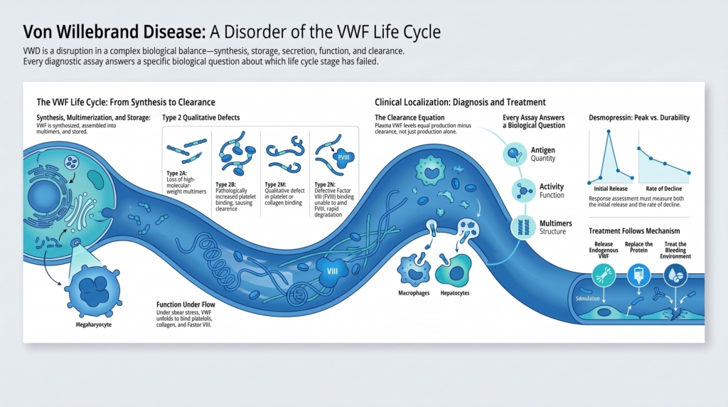
Its distinctive contribution is not simply its breadth. The Primer organizes VWD around the biology and life cycle of von Willebrand factor—from synthesis, multimer assembly, and cellular storage to secretion, function under flow, proteolysis, and clearance—and then connects that biology to phenotype, laboratory diagnosis, classification, genetics, and treatment.
Read it for the map of the field, but especially for the mechanistic framework beneath the map.
What scholarly job does it perform?
Panoramic disease primer
The article constructs a coherent account of:
- epidemiology,
- molecular and cellular biology,
- clinical manifestations,
- diagnostic testing,
- classification,
- genetic diagnosis,
- contemporary management,
- quality of life,
- and future research priorities.
It is not a systematic review answering one narrowly defined question. It is an expert synthesis that decides how the major parts of VWD should fit together.
Review at a glance
| Feature | Assessment |
|---|---|
| Publication year | 2024 |
| Review type | Comprehensive narrative disease primer |
| Best audience | Trainees, general hematologists, laboratory physicians, and specialists seeking an updated overview |
| Best use | Building an integrated mental model of VWD |
| Distinctive contribution | Connects endothelial and vascular VWF biology to clinical diagnosis and treatment |
| Evidence-selection method | Narrative expert review; a reproducible search and study-selection method is not reported |
| Scope exclusions | Acquired von Willebrand syndrome is deliberately excluded; detailed product-level dosing and jurisdiction-specific implementation are not the primary purpose |
| Read with caution | The Primer presents a coherent and authoritative framework, but some of the definitions and treatment positions it adopts remain contested or rest on limited evidence |
Best consulted for
- mechanistic orientation;
- type 1 VWD pathophysiology;
- assay-to-function mapping;
- relationships among synthesis, secretion, clearance, and plasma level;
- broad contemporary diagnosis and management.
Not sufficient alone for
- disputed low-VWF classification;
- detailed perioperative dosing;
- comparative product selection;
- acquired von Willebrand syndrome;
- rapidly changing trial and regulatory information.
Central thesis
The central thesis is that VWD cannot be understood as a single low VWF measurement.
Plasma VWF reflects a sequence of biological processes:
synthesis → dimerization and multimerization → intracellular processing → storage → secretion → unfolding and function under flow → proteolysis → clearance
Disease may arise from disruption at any point in that sequence.
Diagnosis therefore requires more than deciding whether VWF is “low.” It requires integration of:
- the bleeding phenotype,
- VWF antigen,
- platelet-dependent VWF activity,
- factor VIII activity,
- the relationships among those results,
- selectively chosen specialized assays,
- and, in some settings, genetic testing.
Management follows the same principle. Treatment should match not only the diagnostic label but also the mechanism, bleeding challenge, response pattern, and clinical context.
The conceptual model
1. Plasma VWF is governed predominantly by endothelial biology
VWF is synthesized in both endothelial cells and megakaryocytes and is stored in Weibel–Palade bodies and platelet α-granules, respectively. Circulating plasma VWF, however, is governed predominantly by endothelial synthesis, storage, and release.
The review is particularly strong on this intracellular and vascular life cycle.
VWF subunits first dimerize, then assemble into multimers during Golgi processing. The multimers are packaged into endothelial storage organelles and released either constitutively or in response to stimulation. Once secreted, VWF unfolds under flow, participates in platelet adhesion, undergoes ADAMTS13-mediated processing, and is eventually removed from the circulation.
The Primer also gives unusual attention to the clearance machinery. Macrophages, sinusoidal endothelial cells, and hepatocytes participate through receptors including LRP1, macrophage and hepatic lectins, scavenger receptors, stabilin 2, and other glycan-sensitive pathways.
This matters because plasma VWF concentration represents a balance:
what is produced and released versus what is processed and removed
Similar VWF levels may therefore result from very different mechanisms:
- reduced synthesis,
- intracellular retention,
- impaired secretion,
- abnormal multimer processing,
- excessive proteolysis,
- or accelerated clearance.
A VWF result is the net output of a biological system, not a direct measurement of one disease process.
2. Structure explains function—but functions overlap
The Primer links VWF domains to their major interactions:
- D′–D3: factor VIII binding
- A1: platelet GPIb binding and interaction with selected collagen types, particularly collagen IV and VI
- A2: ADAMTS13 cleavage region
- A3: principal binding site for fibrillar collagen, especially collagen I and III
- C4: interaction with platelet integrin αIIbβ3
This domain-based approach makes type 2 VWD easier to understand as a set of disrupted molecular interactions rather than a list of labels.
| Subtype | Principal disturbance |
|---|---|
| 2A | reduced effective high-molecular-weight multimers because of impaired assembly or increased proteolysis |
| 2B | increased affinity of VWF for platelet GPIb |
| 2M | impaired platelet binding or impaired collagen binding—distinct qualitative phenotypes—with preserved multimer distribution |
| 2N | impaired binding and stabilization of factor VIII |
The Primer also acknowledges that the boundaries are not always clean. Some variants produce overlapping phenotypes, particularly across type 2 categories.
3. Diagnosis is relational
The review divides laboratory evaluation into first-level diagnostic assays and second-level classification assays.
First-level assays
- VWF antigen
- platelet-dependent VWF activity
- factor VIII activity
Selectively chosen second-level assays
- multimer analysis
- VWF collagen binding
- ristocetin-induced platelet agglutination
- VWF–factor VIII binding
- VWF propeptide in selected clearance questions
- genetic testing in appropriate cases
The important point is not that every patient proceeds through the same sequence.
Rather:
Specialized testing is selected to resolve the mechanism suggested by the initial pattern.
The relationships among results help localize the defect:
- disproportionately low platelet-dependent activity raises concern for a qualitative platelet-binding or multimer abnormality;
- disproportionately low factor VIII raises concern for defective VWF–factor VIII binding or hemophilia A;
- an increased VWF propeptide-to-antigen ratio can support an accelerated-clearance mechanism.
However, current diagnostic guidance generally favors direct assessment of VWF survival with 1- and 4-hour post-desmopressin measurements when establishing an enhanced-clearance phenotype. VWF propeptide testing can support the mechanism, but it is not the preferred routine substitute for observing the response over time.
The ratio identifies a pattern. It does not complete the diagnosis.
4. Phenotype depends on exposure and time
The review treats bleeding history as indispensable but imperfect.
A patient’s documented phenotype depends partly on whether that patient has encountered:
- menstruation,
- childbirth,
- surgery,
- dental extraction,
- trauma,
- or other hemostatic challenges.
The Primer also acknowledges limitations of bleeding assessment tools:
- reduced sensitivity in people with few prior challenges,
- score saturation when recurrent episodes do not increase the total,
- inability to distinguish VWD from other mild bleeding disorders,
- and dependence on the language, population, and health-care system in which the tool was validated.
The article cites a BAT score above 10 as the best predictor of future treatment-requiring bleeding in one adult cohort. That finding should be understood as study-specific, not as a universal prognostic threshold for every VWD population.
5. Treatment follows mechanism and context
The treatment section can be reduced to three broad strategies.
Release endogenous VWF
Desmopressin
Useful when sufficient functional VWF can be released and when the response is both adequate and durable.
Replace deficient or dysfunctional VWF
Plasma-derived or recombinant VWF
Used when desmopressin is ineffective, contraindicated, or insufficient for the clinical challenge.
Treat the bleeding environment
Antifibrinolytics, hormonal therapy, local hemostatic measures, iron replacement, and lesion-directed interventions
This matters because many VWD bleeds occur at mucosal or vascular surfaces where raising VWF alone may not solve the entire problem.
What this review uniquely offers
1. A life-cycle view of VWD
Many reviews describe VWF structure and treatment. This one more effectively connects:
- endothelial and megakaryocyte synthesis,
- dimerization and multimerization,
- Weibel–Palade body storage,
- stimulated secretion,
- shear-dependent unfolding,
- ADAMTS13 processing,
- glycosylation,
- and receptor-mediated clearance.
The detail devoted to the clearance-receptor network is especially useful. It shows how LRP1, macrophage and hepatic lectins, scavenger receptors, stabilin 2, and related pathways help determine how long VWF remains in the circulation.
That framework is particularly valuable for understanding type 1 disease and low VWF, where the same plasma concentration may arise from different biological processes.
2. A mechanistic account of type 1 VWD
The review explicitly recognizes that type 1 VWD may result from:
- reduced synthesis,
- defective intracellular storage,
- impaired secretion,
- accelerated clearance,
- or mechanisms not directly attributable to a pathogenic variant in the VWF gene.
That last point is important. It helps explain why a patient can have a convincing quantitative phenotype without a clearly pathogenic VWF variant.
3. Clear linkage between assays and biological questions
The review does not merely list tests. It shows that each assay interrogates a different question:
- How much VWF is present?
- Does it bind platelets?
- Does it bind collagen?
- Are high-molecular-weight multimers preserved?
- Does it bind and stabilize factor VIII?
- Does it remain in the circulation normally?
4. Integration of disease burden
Heavy menstrual bleeding, iron deficiency, joint bleeding, treatment burden, and quality of life are treated as central clinical concerns rather than secondary topics.
Iron deficiency is appropriately understood as an important consequence and marker of cumulative bleeding burden, not as a bleeding manifestation itself.
Where interpretation begins
An expert review does not merely reproduce evidence. It selects definitions, resolves ambiguities, and advances a particular view of the field.
Three positions are especially important in this Primer.
1. It adopts the 2021 classification of symptomatic low VWF as type 1 VWD
The authors explicitly use the 2021 ASH/ISTH/NHF/WFH framework in which patients with VWF levels of 30–50 IU/dL and abnormal bleeding may be diagnosed with type 1 VWD.
This is not an incidental wording choice. It places the review on one side of a continuing debate.
The position may improve diagnostic access and avoid withholding care from symptomatic patients. But competing guidance and newer literature continue to ask whether everyone within this range shares the same disease biology, inheritance pattern, or long-term bleeding risk.
Several authors of the Primer helped shape the contemporary diagnostic framework. The article therefore represents one of the most authoritative integrated statements of the broader type 1 position—but not a neutral account of all sides of the dispute.
Its authority and its partisanship are two aspects of the same fact.
2. It incorporates the formally recognized but operationally evolving type 1C category
The Primer incorporates type 1C for patients with accelerated VWF clearance.
This has a direct clinical implication. In an enhanced-clearance phenotype, the response to desmopressin may appear adequate at 1 hour but fall substantially by 4 hours. Measuring only the early peak can therefore misclassify the patient as a durable responder.
Type 1C is formally recognized, but its practical definition, optimal diagnostic strategy, and frequency of routine assignment remain evolving.
The category may be best understood as both:
- a mechanistic subtype, and
- a pharmacokinetic phenotype with direct implications for treatment planning.
3. It argues that type 1 VWD can arise independently of the VWF gene
The review emphasizes that many patients with VWF levels of 30–50 IU/dL do not have an identifiable pathogenic VWF variant and that linkage and population studies support contributions from other genetic loci.
This moves type 1 VWD away from a simple one-gene, one-disease model.
It also explains why genotype may clarify selected cases without serving as a universal diagnostic arbiter.
What it decides for the reader
The Primer provides a defensible working framework:
- understand VWF as a dynamic biological system rather than a static level;
- interpret antigen, activity, and factor VIII in relation to one another;
- choose specialized tests according to the suspected defect;
- assess desmopressin response for both magnitude and durability;
- use type 2 classification when it localizes a functional defect and changes management;
- use genetic testing selectively, especially in type 2 and type 3 disease;
- seek iron deficiency actively in patients with chronic mucosal or menstrual bleeding;
- combine VWF-directed therapy with site-specific and supportive care.
What it leaves open
The Primer identifies—but does not fully resolve—several central questions:
- Should all symptomatic patients with VWF levels of 30–50 IU/dL be classified as having type 1 VWD?
- How should clinicians manage patients with borderline levels but limited prior hemostatic exposure?
- When VWF rises with age, does bleeding risk normalize as well?
- How should overlapping type 2 phenotypes be classified?
- How often should type 1C be formally assigned rather than described as an enhanced-clearance phenotype?
- Which patients derive the greatest benefit from prophylaxis?
- What dosing, trough targets, and stopping rules should guide prophylaxis?
- Which emerging therapies will improve patient-important bleeding outcomes rather than laboratory measurements alone?
These are places where readers must return to focused reviews, original investigations, and guidelines.
What has changed since publication?
The Primer was published in 2024 and remains contemporary, but several sections will age faster than others.
Areas requiring periodic reassessment include:
- the classification of low VWF and mild type 1 disease;
- operational use of type 1C;
- qualitative low-VWF phenotypes;
- assay platforms and laboratory thresholds;
- prophylaxis evidence;
- age-related changes in VWF;
- new molecular and polygenic models;
- emerging therapies;
- and trial or regulatory status.
Strengths
- Integrates basic biology with clinical care.
- Provides unusually strong figures, particularly for the VWF life cycle, type 2 mechanisms, diagnostic localization, and genotype–domain relationships.
- Explains quantitative disease mechanistically, rather than treating type 1 VWD as a homogeneous reduction in protein level.
- Connects laboratory assays to biological functions.
- Includes quality of life, menstrual bleeding, iron deficiency, prophylaxis, and future therapies.
- Identifies major research gaps rather than presenting VWD as a settled field.
Limitations
- It is a narrative expert synthesis, without a reported reproducible evidence-search or selection method.
- Its breadth limits the depth devoted to individual controversies.
- The diagnostic framework largely reflects the 2021 international guidelines and the views of authors closely involved in modern VWD classification. Readers seeking direct challenges to that framework will need other sources.
- Guideline recommendations, mechanistic evidence, observational studies, and expert judgment sometimes appear close together, making differences in evidentiary certainty less visible.
- Evidence remains limited for several treatment areas, including prophylaxis, gastrointestinal bleeding, pregnancy, alloantibodies, and novel therapies.
- Acquired von Willebrand syndrome is excluded.
- The article is dense and is more useful as a section-based reference than as the first VWD text for a novice learner.
- Several authors report industry relationships, research support, advisory roles, or patent interests relevant to VWF diagnostics and therapy. These disclosures deserve attention, particularly in the treatment and future-therapy sections.
How to read this review
First pass: Build the map
Read:
- the abstract and section headings;
- Figure 2, showing the VWF life cycle;
- Figure 4, showing type 2 mechanisms;
- Figure 6, showing the diagnostic algorithm;
- the Outlook section.
This establishes the paper’s architecture before the details accumulate.
Second pass: Follow one mechanistic thread
Choose one pathway, for example:
synthesis → secretion → plasma survival → laboratory pattern → desmopressin response
This is particularly useful for understanding type 1 and enhanced-clearance phenotypes.
Third pass: Follow one clinical problem
Choose:
- suspected type 2 VWD,
- low VWF,
- heavy menstrual bleeding,
- gastrointestinal bleeding,
- pregnancy,
- or recurrent bleeding requiring prophylaxis.
Then trace how the authors move from mechanism to diagnosis to management.
Fourth pass: Mark the judgments
Look for places where the authors:
- adopt a disputed definition,
- favor a diagnostic threshold,
- endorse a classification,
- advocate broader treatment,
- or extrapolate from mechanistic evidence.
Those passages reveal the review’s point of view.
Clinical pearls
- A VWF level is a snapshot of a dynamic biological system.
- Antigen measures quantity; activity assays interrogate selected functions.
- Ratios localize discordance but do not establish the final diagnosis alone.
- Normal results obtained during acute bleeding, physiological stress, inflammation, pregnancy, or another recognized VWF-elevating state may require reassessment when clinical suspicion remains high.
- The recorded bleeding phenotype depends partly on prior hemostatic opportunity.
- Desmopressin response must be evaluated for both peak and durability.
- An adequate 1-hour response with substantial decline by 4 hours should raise concern for accelerated clearance.
- Iron deficiency is an important consequence and marker of cumulative bleeding burden.
- Type 2N VWD and mild hemophilia A must be distinguished because treatment and inheritance differ.
- Gastrointestinal bleeding may reflect both impaired hemostasis and angiodysplasia.
- Prophylaxis should be individualized and periodically reassessed rather than assigned solely by VWD subtype.
Where it fits in the VWD module
This should serve as the panoramic anchor for the Review Library.
It connects most directly to:
- VWF in One Picture
- Classification as a Tool
- Diagnostic Reasoning as Localization
- Interpreting VWF Labs Relationally
- The Diagnostic Test Atlas
- Normal VWF Variation versus Disease
- Desmopressin Trial
- VWF Replacement Therapy
- Long-Term Prophylaxis
- Heavy Menstrual Bleeding and Iron Deficiency
- Pregnancy and Delivery
- Future Therapies
It should not replace those essays. The Primer supplies the integrated map; the module’s individual spokes examine the difficult regions of that map in greater depth.
Read next
To challenge the diagnostic framework
Brenner MK, Christopherson PA, Flood VH. (Un)Diagnosing von Willebrand disease.
Read this for a more explicit examination of overdiagnosis, label instability, reassessment, and diagnostic thresholds.
To explore the low-VWF controversy
O’Donnell JS. Low von Willebrand factor—unraveling an enigma wrapped in a conundrum.
Read this as an argument about heterogeneous biology, bleeding opportunity, and the risks of treating a laboratory range as a single disease state.
To see how the field changed
Leebeek FWG, Eikenboom JCJ. Von Willebrand’s disease. New England Journal of Medicine. 2016.
Read this as an earlier benchmark and compare its treatment of classification, assays, genetics, prophylaxis, and low VWF with the 2024 Primer.
Bottom line
The 2024 Nature Reviews Disease Primers article is one of the strongest contemporary single-paper orientations to VWD.
What makes it especially valuable is not simply its comprehensiveness. It places the VWF life cycle and endothelial biology of circulating VWF at the center of the disease and uses that framework to connect plasma levels, functional defects, genotype, laboratory testing, desmopressin response, and emerging therapy.
But it is not a neutral survey. It adopts the 2021 low-VWF classification, incorporates the formally recognized but still operationally evolving type 1C category, and advances a broader genetic model of type 1 VWD. Those positions are useful precisely because they can be identified, examined, and compared with other reviews.
Read it for the map—and then read more focused literature where the map’s borders remain disputed.
Reflect and discuss
- Does classifying symptomatic VWF levels of 30–50 IU/dL as type 1 VWD improve care, or combine biologically different states under one label?
- Does type 1C represent a distinct disease subtype, a pharmacokinetic phenotype, or both?
- Which parts of the diagnostic algorithm reflect biological localization, and which depend primarily on negotiated thresholds?
- If VWF levels normalize with age, what must be shown before concluding that the bleeding disorder has also resolved?
- Where does the Primer report established evidence, and where does it advance a preferred interpretation of that evidence?