
Why VWD bleeding is often mucosal, recurrent, and challenge-dependent
Note: The video and audio linked above were generated with the assistance of AI. Clinical accuracy has been reviewed, but no AI-generated content can be guaranteed to be fully error-free.
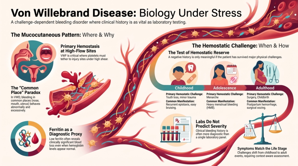
Why this spoke matters
Von Willebrand disease is often described as a mucocutaneous bleeding disorder.
That description is accurate.
But it is not enough.
The clinical presentation of VWD reflects the biology of von Willebrand factor itself. VWF supports platelet adhesion at sites of vascular injury, especially under shear; helps stabilize factor VIII; and becomes most important when local hemostatic demand exceeds baseline reserve.1
That is why many patients with VWD do not bleed every day.
They bleed when challenged.
A child may present with recurrent epistaxis or easy bruising. An adolescent may present with heavy menstrual bleeding. An adult may present after dental extraction, surgery, childbirth, or recurrent iron deficiency. An older patient with severe VWD, high-molecular-weight multimer loss, or acquired von Willebrand syndrome may present with gastrointestinal bleeding from angiodysplasia.2
The question is not simply:
Does this patient bleed?
The better question is:
When has this patient’s hemostatic system been tested, and what happened when it was?
The core pattern
The typical bleeding pattern in VWD is mucocutaneous.
That means bleeding from surfaces and tissues where primary hemostasis is especially important, including the nose, mouth, gums, skin, uterus, gastrointestinal mucosa, dental sites, and sites of mucosal or surgical injury.
This pattern reflects the role of VWF in platelet adhesion and early platelet plug formation. When VWF quantity or function is reduced, the initial platelet response to vascular injury may be inefficient, especially in vascular beds exposed to shear, local fibrinolysis, mechanical stress, or repeated minor trauma.3
This is why VWD often presents with:
- epistaxis
- easy bruising
- gum or oral bleeding
- prolonged bleeding from minor wounds
- heavy menstrual bleeding
- postpartum bleeding
- bleeding after dental extraction
- bleeding after surgery or procedures
The pattern is suggestive, but it is not diagnostic by itself. Bruising, epistaxis, and heavy menstrual bleeding are common in the general population. What matters is severity, recurrence, treatment requirement, association with iron deficiency or transfusion, family history, laboratory pattern, and response to hemostatic challenge.4
The pattern is not random.
It is VWF biology made visible.
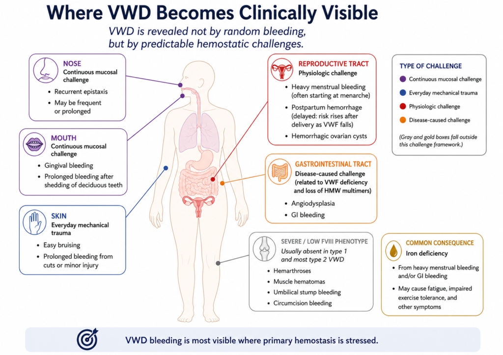
Why mucosal surfaces reveal VWD
Mucosal surfaces are hemostatically demanding.
They are vascular. They are exposed to friction, local trauma, microbial inflammation, and fibrinolytic activity. They also bleed in places where pressure and immobilization may be difficult to achieve.
VWF is especially important in these settings because it helps platelets tether to sites of injury under flow.
When VWF is low, dysfunctional, lacking high-molecular-weight multimers, cleared too quickly, or unable to support normal platelet adhesion, mucosal bleeding becomes more likely.
This explains why VWD often presents as “ordinary” bleeding that becomes excessive:
a nosebleed that lasts too long
a period that is heavier than expected
bruises that are larger or more frequent than expected
bleeding after dental work that does not stop on schedule
postpartum bleeding that exceeds expected physiologic blood loss
In VWD, the problem is often not that bleeding starts in strange places.
It is that bleeding in common places behaves abnormally.
Bleeding is often challenge-dependent
Many patients with VWD look well between hemostatic challenges.
This can be misleading.
A patient may not bleed every day because everyday life has not recently tested VWF-dependent hemostasis. The bleeding history becomes meaningful only when interpreted in relation to exposure.
Important hemostatic challenges include dental extraction, surgery, menarche, childbirth, tonsillectomy, trauma, recurrent mucosal inflammation, and exposure to anticoagulant or antiplatelet therapy.
A negative bleeding history is more reassuring when the patient has passed meaningful hemostatic challenges without bleeding. It is less reassuring when the patient has never been challenged.
This is especially important in children, adolescents before menarche, men without prior procedures, and adults who have avoided surgery.5
The absence of bleeding may mean normal hemostasis.
Or it may mean that the test has not yet occurred.
Bleeding across the lifespan
VWD often presents differently at different ages.
In childhood, the first clues may be recurrent epistaxis, easy bruising, oral bleeding, or bleeding after tonsillectomy, dental work, immunizations, loss of teeth, or minor procedures.
But childhood bleeding is difficult to interpret. Bruising and nosebleeds are common in children without bleeding disorders. Children may also have had few major hemostatic challenges. A low bleeding score in a young child is therefore not as reassuring as a low bleeding score in an adult who has had surgery, dental extraction, childbirth, or trauma without bleeding.6
In adolescence, heavy menstrual bleeding may become the first major hemostatic challenge.
In adulthood, VWD may appear through surgical bleeding, postpartum hemorrhage, recurrent iron deficiency, or delayed recognition of lifelong mucocutaneous bleeding.
In older adults, the presentation may shift again. Gastrointestinal bleeding, angiodysplasia, comorbid illness, medications, and acquired von Willebrand syndrome may complicate the picture.
The disease is the same diagnostic family.
But the hemostatic challenges change over time.
Heavy menstrual bleeding and iron deficiency
Heavy menstrual bleeding is one of the most important clinical manifestations of VWD.
It may present as prolonged menses, flooding, passage of clots, frequent pad or tampon changes, nighttime changes, missed school or work, iron deficiency, or iron deficiency anemia.
Heavy menstrual bleeding is not just a quality-of-life issue.
It is often the clinical event that reveals reduced hemostatic reserve.
In patients who menstruate, the uterus may become the first repeated, high-demand test of primary hemostasis. A patient who has had few childhood bleeding symptoms may develop clinically obvious bleeding at menarche or in adolescence.
Practical clues to clinically significant menstrual blood loss include clots larger than about 1 inch, changing a pad or tampon more than hourly, and low ferritin.7
Iron deficiency is a key part of the presentation. A normal hemoglobin does not exclude clinically important iron loss, particularly in patients with chronic heavy menstrual or mucosal bleeding.8
For this reason, evaluation of suspected VWD in a patient with heavy menstrual bleeding should include attention to iron stores, not only hemoglobin.
Ferritin is part of the bleeding story.
Epistaxis, bruising, and oral bleeding
Epistaxis is common in childhood, including in children without bleeding disorders.
That creates a diagnostic challenge.
The question is not simply whether nosebleeds occur, but whether they are excessive for age and context. Frequency, duration, bilateral bleeding, need for medical attention, packing, cautery, iron deficiency, and family history all matter.
Easy bruising is similar. Bruising is common in active children. But large bruises, spontaneous bruises, bruises in unusual locations, bruising out of proportion to trauma, bruising with a palpable lump, or bruising associated with other mucosal bleeding may raise suspicion.9
Oral bleeding is especially informative because the mouth is a demanding hemostatic environment. Gingival bleeding, prolonged bleeding after dental cleaning, bleeding after tooth loss, and especially bleeding after dental extraction may reveal VWF-dependent vulnerability.
Dental extraction combines vascularity, tissue disruption, saliva, local fibrinolysis, and mechanical stress.
When bleeding after dental work is excessive, recurrent, or delayed, it should be taken seriously.
Surgery, procedures, and recurrent bleeding
VWD may first become obvious after surgery.
The bleeding may be immediate, delayed, recurrent, or persistent.
This matters because VWD bleeding is often not a dramatic intraoperative hemorrhage. It may appear as persistent oozing, recurrent wound bleeding, oral bleeding after tonsillectomy or dental extraction, or bleeding after a procedure that was expected to be minor.
Mucosal procedures are especially important:
tonsillectomy
adenoidectomy
dental extraction
nasal surgery
gynecologic procedures
gastrointestinal procedures with biopsy or polypectomy
Tonsillectomy deserves special caution because the airway is involved and bleeding may occur later in the postoperative course as the throat heals. In a patient with suspected or confirmed VWD, perioperative planning may involve antifibrinolytic therapy, desmopressin if appropriate and documented to be effective, VWF replacement in selected cases, and individualized target monitoring depending on the procedure and subtype.10
Delayed or recurrent bleeding can occur after tonsillectomy, dental extraction, childbirth, or other mucosal procedures and should not be dismissed because initial hemostasis appeared satisfactory.
A surgical history is therefore not just a list of operations.
It is a record of how the patient handled hemostatic stress.
Pregnancy, delivery, and postpartum bleeding
Pregnancy is a dynamic hemostatic state.
In many patients with type 1 VWD, VWF and factor VIII levels rise during pregnancy. This may improve hemostatic reserve near delivery. But the rise is variable, may be inadequate, and does not necessarily protect against postpartum bleeding.11
The postpartum period is especially important because VWF and factor VIII levels fall after delivery. Bleeding may occur after the immediate delivery window, especially as pregnancy-associated hemostatic changes recede.12
VWD may present as:
- primary postpartum hemorrhage
- secondary postpartum hemorrhage
- bleeding after miscarriage or pregnancy procedures
- iron deficiency after delivery
- heavy postpartum lochia
- need for transfusion or hemostatic therapy
Pregnancy also reveals the limits of interpreting a single VWF level. A normal VWF level during pregnancy does not necessarily represent the patient’s baseline.
Timing matters.
So does the story.
Gastrointestinal bleeding and angiodysplasia
Gastrointestinal bleeding is not the most common presentation of VWD overall, but it is clinically important.
It is especially associated with severe VWD, type 3 VWD, and qualitative or acquired states involving loss of high-molecular-weight multimers, including type 2A-like patterns and acquired von Willebrand syndrome.
Angiodysplasia is a key association.
The biology is not simply “low clotting factor.” VWF appears to contribute both to hemostasis at high-shear mucosal vascular lesions and to vascular biology more broadly. Loss of high-molecular-weight multimers can impair platelet adhesion in fragile mucosal vascular beds, increasing risk of recurrent gastrointestinal bleeding.13
Recurrent gastrointestinal bleeding can be difficult to manage and may require attention not only to hemostatic replacement, but also to the underlying vascular lesion and to acquired causes.
In an older patient with new gastrointestinal bleeding, especially with aortic stenosis, LVAD support, plasma cell dyscrasia, lymphoproliferative disease, marked thrombocytosis or myeloproliferative neoplasm, hypothyroidism, or no lifelong bleeding history, acquired von Willebrand syndrome should be considered.14
Not all VWF-mediated bleeding is inherited.
When VWD looks hemophilia-like
Most VWD bleeding is mucocutaneous.
But severe VWD can look more like a coagulation factor deficiency.
This occurs because VWF stabilizes factor VIII. When VWF is nearly absent, as in type 3 VWD, factor VIII levels may be very low. Patients may develop deep tissue bleeding, muscle hematomas, hemarthroses, and a more hemophilia-like phenotype.
Type 2N VWD can also resemble hemophilia A because VWF cannot bind and stabilize factor VIII normally. The patient may have disproportionately low factor VIII with VWF antigen and platelet-dependent activity that are not profoundly reduced.15
This is clinically important because a low factor VIII level should not automatically be assumed to mean hemophilia A.
Sometimes the problem is not factor VIII production.
Sometimes the problem is VWF failing to carry factor VIII.
Why bleeding severity does not always match the VWF level
VWF level matters.
But it does not fully predict bleeding.
Two patients with similar VWF levels may have different bleeding histories. One may have heavy menstrual bleeding, postpartum hemorrhage, and dental bleeding. Another may have few symptoms despite similar laboratory values.
Several factors help explain this mismatch:
- VWD subtype
- VWF activity relative to antigen
- multimer pattern
- factor VIII level
- age
- ABO blood group
- hormonal state
- inflammation
- prior hemostatic challenges
- local anatomy
- medications
- platelet function
- iron deficiency
- comorbid illness
Bleeding assessment tools can help organize the history, but they have limitations. They depend on age, recall, sex, cumulative scoring, and prior opportunities to bleed. A low score may be falsely reassuring in someone who has not yet had major hemostatic challenges.16
BATs are most useful as structured screening tools in low-prevalence settings and as documentation tools in referral settings. They should not be used alone to decide against testing when clinical suspicion is meaningful or a first-degree relative is affected.17
Bleeding is not a laboratory value.
It is the clinical result of a hemostatic system meeting real-world challenges.
That is why the bleeding history is not secondary to the lab.
It is part of the diagnosis.
Normal screening tests do not exclude VWD
The clinical manifestations of VWD may be present even when routine screening tests are normal.
A normal platelet count does not exclude VWD. The PT is typically normal. A normal aPTT does not exclude VWD because factor VIII may be normal or only mildly reduced, especially in mild type 1 disease and many qualitative subtypes.
Likewise, global screening tools such as PFA-100/200 can be abnormal in VWD, especially in severe disease or some qualitative subtypes, but they are not reliable enough to rule out VWD when the clinical story is persuasive.18
When the bleeding phenotype is convincing, the appropriate next step is not reassurance based on normal screening tests.
It is focused VWF testing interpreted in clinical context.
Clinical synthesis
The clinical manifestations of VWD reflect the biology of VWF.
VWF is most important where platelet adhesion must occur quickly, under flow, and at vulnerable mucosal surfaces. It also stabilizes factor VIII, which explains why severe VWF deficiency can produce deep bleeding that resembles hemophilia.
But the absence of daily bleeding does not exclude VWD, and the presence of common bleeding symptoms does not establish it.
The bleeding history must be read against exposure: what challenges occurred, how demanding they were, and what happened afterward.
In VWD, clinical presentation is best understood as the interaction between VWF biology, hemostatic challenge, vascular bed, and time.
Evidence anchor: why VWD bleeding is mucosal, variable, and challenge-dependent
Summary derived from clinical cohorts, diagnostic guidelines, women’s health literature, pediatric reviews, subtype studies, and laboratory guidance. The evidence consistently shows that VWD bleeding is common at mucosal surfaces, varies across the lifespan, and cannot be predicted from a single symptom or VWF value alone.
| Evidence stream | What it shows | Why it matters | Main limitation |
|---|---|---|---|
| Clinical phenotype | VWD commonly presents with epistaxis, bruising, oral bleeding, heavy menstrual bleeding, postpartum bleeding, bleeding from minor wounds, and bleeding after dental work, surgery, or childbirth.19 | The clinical pattern reflects impaired primary hemostasis at mucosal and procedural sites. | Symptom frequency varies by subtype, age, sex, referral setting, and prior hemostatic challenges. |
| General-population symptom overlap | Bruising, epistaxis, and heavy menstrual bleeding are also common in people without VWD.20 | Symptoms must be interpreted by severity, recurrence, treatment requirement, family history, laboratory pattern, and challenge exposure. | Common symptoms can be overinterpreted when laboratory values are borderline. |
| Women’s health | Heavy menstrual bleeding and postpartum bleeding are major manifestations of VWD, and iron deficiency may be an important consequence of chronic menstrual blood loss.21 | VWD may first become clinically obvious at menarche, childbirth, or through iron deficiency. | Heavy menstrual bleeding is common and has gynecologic as well as hematologic causes. |
| Pediatric presentation | Bruising and epistaxis are common in children with VWD, but they are also common in children without bleeding disorders; many children have not yet had major hemostatic challenges.22 | Pediatric bleeding histories require context, not reflexive labeling. | A low bleeding score may be falsely reassuring when the child has not yet been challenged. |
| Subtype effects | Severe type 3 VWD and type 2N VWD may produce low FVIII and deep tissue, muscle, or joint bleeding, creating a hemophilia-like phenotype.23 | VWD can sometimes resemble hemophilia when FVIII stabilization is impaired. | FVIII level contributes to bleeding phenotype but does not fully determine it. |
| GI bleeding and angiodysplasia | Gastrointestinal bleeding and angiodysplasia are enriched in severe VWD, high-molecular-weight multimer-loss states, and acquired VWS.24 | Recurrent GI bleeding should prompt attention to multimer biology, vascular lesions, and acquired causes. | GI bleeding has many non-VWD causes, especially in older adults. |
| Bleeding assessment tools | BATs help standardize bleeding history, but their interpretation depends on age, sex, recall, cumulative scoring, and prior hemostatic challenges.25 | BATs are useful for structure and documentation, but they do not replace clinical judgment. | BATs may underperform in patients who have not yet had meaningful bleeding challenges. |
| Laboratory guidance | Normal PT, aPTT, platelet count, or PFA-100/200 testing cannot reliably exclude VWD when the clinical pattern is persuasive.26 | Focused VWF testing is needed when the bleeding history suggests VWF-mediated bleeding. | VWF testing itself is sensitive to preanalytic conditions, physiologic modifiers, assay choice, and repeat testing context. |
Interpretive note: The central clinical question is whether the patient has bled excessively for the hemostatic challenges they have faced, and whether the pattern fits clinically meaningful VWF-mediated bleeding.
Clinical guidance: reading the bleeding history in suspected VWD
Do not ask only, “Does the patient bleed?”
Ask what kind of bleeding, in what setting, and after what challenge.
Look for mucocutaneous bleeding
Epistaxis, easy bruising, gum bleeding, oral bleeding, heavy menstrual bleeding, postpartum bleeding, and bleeding after dental extraction or mucosal procedures are especially relevant.
Ask about hemostatic challenges
A negative bleeding history is more meaningful after dental extraction, surgery, childbirth, trauma, tonsillectomy, or other significant challenges. It is less meaningful when those challenges have not occurred.
Treat heavy menstrual bleeding as hematologic evidence
Ask about flooding, clots, duration, frequency of product changes, nighttime changes, missed school or work, iron deficiency, anemia, and response to hormonal or hemostatic therapy. Clots larger than about 1 inch, changing a pad or tampon more than hourly, and low ferritin are practical clues to clinically significant menstrual blood loss.
Ask about delayed or recurrent bleeding
Bleeding after tonsillectomy, dental extraction, childbirth, or mucosal procedures may occur after the initial procedure. Delayed or recurrent bleeding should not be dismissed because the first hours looked reassuring.
Check iron stores
Iron deficiency may be the measurable consequence of chronic mucosal bleeding. Ferritin can reveal clinically important blood loss even when hemoglobin is preserved.
Interpret pediatric bleeding carefully
Bruising and epistaxis are common in children. A bleeding history becomes more persuasive when bleeding is recurrent, severe, treatment-requiring, associated with iron deficiency, supported by family history, or occurs after a meaningful hemostatic challenge.
Use BATs thoughtfully
Bleeding assessment tools can standardize and document the bleeding history. They are most useful as screening tools in low-prevalence settings and as severity tools in referral settings. They should not be used alone to decide against testing when clinical suspicion is meaningful or a first-degree relative is affected.
Do not overtrust normal screening tests
A normal platelet count, PT, aPTT, or PFA-100/200 does not exclude VWD. If the bleeding pattern is convincing, order VWF antigen, platelet-dependent VWF activity, and factor VIII activity, with repeat testing as needed.
Consider subtype and acquired causes
Deep tissue bleeding, joint bleeding, very low factor VIII, recurrent gastrointestinal bleeding, angiodysplasia, older age, aortic stenosis, LVAD support, plasma cell dyscrasia, lymphoproliferative disease, marked thrombocytosis or myeloproliferative neoplasm, hypothyroidism, or new bleeding without a lifelong history should broaden the differential.
Look for alternate or coexisting explanations
Consider thrombocytopenia, platelet dysfunction, liver disease, kidney disease, connective tissue disorders, telangiectasia, gynecologic pathology, medications, and acquired von Willebrand syndrome.
Interpret the story with the laboratory pattern
The bleeding phenotype, VWF antigen, platelet-dependent activity, factor VIII, activity-to-antigen ratio, multimer pattern, collagen binding, family history, and repeat testing should be interpreted together.
Reflect & Apply Case
A 16-year-old girl is referred for heavy menstrual bleeding and iron deficiency.
She reports menses lasting 8 to 10 days, flooding on the heaviest days, passage of clots, and changing protection every 1 to 2 hours. She has missed school because of bleeding. Her ferritin is 8 ng/mL, but her hemoglobin is only mildly reduced.
She also reports easy bruising and occasional nosebleeds. She has never had surgery, dental extraction, pregnancy, or major trauma.
Initial VWD testing shows:
- VWF antigen: 44 IU/dL
- platelet-dependent VWF activity: 41 IU/dL
- factor VIII activity: 52 IU/dL
- activity-to-antigen ratio: preserved
- platelet count: normal
Questions for reflection:
How much weight should you give the heavy menstrual bleeding?
How does iron deficiency change your interpretation?
Does the absence of surgical bleeding reassure you if she has never had surgery?
What additional bleeding history would you ask for?
Would a normal PT or aPTT exclude VWD?
How would you interpret a normal bleeding score in someone with few prior hemostatic challenges?
What repeat testing or additional assays might be useful?
How would you plan for a future dental extraction or procedure?
The point is not to decide from one laboratory panel alone.
The point is to recognize that her uterus may already have served as a major hemostatic challenge.
In VWD, bleeding history is not just background information.
It is biology under stress.
Test your thinking
An interactive quiz for this topic is being developed and will be added soon.