
Why VWD types are best understood as VWF failure modes
Note: The video and audio linked above were generated with the assistance of AI. Clinical accuracy has been reviewed, but no AI-generated content can be guaranteed to be fully error-free.
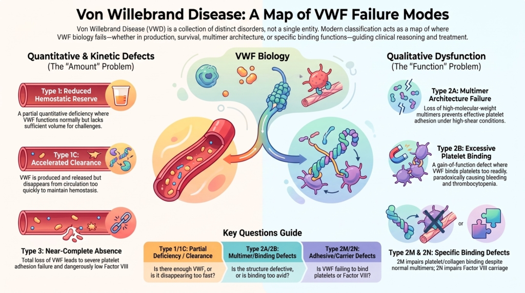
Why this spoke matters
VWD is often introduced as a list: type 1, type 2A, type 2B, type 2M, type 2N, and type 3.
For many learners, classification becomes an exercise in memorization.
But the classification system did not emerge arbitrarily.
It evolved because different abnormalities in VWF biology produce different patterns of bleeding, different laboratory signatures, and different therapeutic implications.
The first step is knowing that von Willebrand disease is not one disease.
The next step is understanding why its categories behave differently.
VWD classification is not simply a naming system. At its best, it is a map of VWF failure modes. The label is useful only when it points beyond itself to the mechanism underneath.
The best question is not simply:
Which subtype is this?
The better question is:
What aspect of VWF biology is failing?
Why classification works
VWF has several jobs.
It must be produced, multimerized, stored, secreted, preserved in circulation, tether platelets under shear, interact with collagen, and stabilize factor VIII. Defects at different points in that biology produce different clinical and laboratory patterns.
That is the logic behind the modern classification of VWD.
Modern classification separates quantitative deficiency from qualitative dysfunction and then localizes the dominant VWF failure mode.1
This system is not perfect. No clinical classification is. But in VWD, many categories correspond remarkably well to recognizable VWF failure modes.
This is especially true for the type 2 subtypes, where the label often identifies the dominant functional defect rather than merely describing severity.
VWD classification is therefore more than severity grading. It is a map of where VWF biology fails: quantity, survival, multimer architecture, platelet binding, collagen binding, factor VIII binding, or near-complete absence.
That makes classification clinically useful.
It helps clinicians interpret laboratory patterns, anticipate treatment response, choose additional tests, plan procedures, and counsel families.
Quantitative and qualitative disorders
Modern VWD classification begins with a broad distinction between quantitative disorders and qualitative disorders.
| Major category | Core abnormality | Fundamental problem |
|---|---|---|
| Quantitative disorders | Reduced amount of VWF | Insufficient functional protein |
| Qualitative disorders | Dysfunctional VWF | Protein present but functionally abnormal |
In quantitative disorders, the major problem is insufficient VWF. The molecule is generally functionally intact, but there is not enough of it.
In qualitative disorders, VWF may be present in normal or near-normal amounts, but one or more key functions of the molecule are impaired. Depending on the subtype, those defects may involve multimer assembly, platelet interaction, collagen binding, or factor VIII stabilization.
This framework shifts the question from “How much VWF is present?” to “How is the molecule behaving?”
A structure-function overview of VWD subtypes
The categories become easier to remember when they are taught as failure modes, not as isolated labels.
| Type | Core defect | Main biologic failure | Typical laboratory pattern | Major clinical implication |
| Type 1 | Partial quantitative deficiency | Reduced hemostatic reserve | Proportional reduction in VWF antigen and activity | Variable mucocutaneous bleeding, often challenge-dependent |
| Type 1C | Accelerated clearance | Shortened VWF survival | Brief or poorly sustained DDAVP response; sometimes elevated VWFpp:VWF:Ag ratio | Baseline level may underestimate kinetic problem |
| Type 3 | Near-complete VWF deficiency | Loss of multiple VWF functions | Very low or absent VWF; low FVIII | Severe bleeding, sometimes hemophilia-like |
| Type 2A | Loss of high-molecular-weight multimers | Impaired platelet adhesion under shear | Reduced HMW multimers; low activity-to-antigen ratio | Mucocutaneous bleeding; impaired high-shear hemostasis |
| Type 2B | Increased platelet binding | Pathologic VWF-platelet interaction and clearance | HMW multimer loss, enhanced platelet binding, possible thrombocytopenia | Thrombocytopenia may worsen with stress or DDAVP |
| Type 2M | Functional adhesive defect | Dysfunction despite preserved multimers | Disproportionately low platelet-dependent activity or collagen binding with relatively preserved multimers | Can be missed if multimers alone are overtrusted |
| Type 2N | Impaired FVIII binding | Reduced FVIII stabilization | Low FVIII disproportionate to VWF antigen and activity | Hemophilia A mimic; usually recessive |
| Low VWF | Borderline quantitative reduction | Reduced reserve with variable penetrance | Mild quantitative reduction | Term retained in clinical usage, although it is no longer a separate diagnostic category in the 2021 guideline |
Sources:2
Type 1 VWD: reduced hemostatic reserve
Type 1 VWD is a partial quantitative deficiency of VWF.
The protein is present, and it usually functions relatively normally, but there is not enough of it. Because both VWF antigen and platelet-dependent VWF activity are reduced proportionally, the activity-to-antigen ratio is often preserved. Multimers are usually normal.
The clinical idea is reduced hemostatic reserve.
Many patients do not bleed continuously. They bleed when a hemostatic challenge exceeds the reserve available to them: menstruation, childbirth, dental extraction, surgery, trauma, or mucosal injury.
That is why type 1 VWD can be both real and variable. A patient may appear well in ordinary life but declare the disorder during a challenge.
The mechanism is quantitative, but the phenotype is contextual.
Type 1C VWD: when survival is the problem
Some patients have a type 1-like pattern because VWF is cleared too quickly.
This is often called type 1C, where “C” refers to clearance. The patient may produce and release VWF, but the protein does not persist normally in circulation.3
This distinction matters because the baseline VWF level does not tell the whole story.
Two patients may have similar VWF levels. One may make too little VWF. The other may release VWF but clear it rapidly. Their laboratory values may look similar at rest, but their response to desmopressin may differ over time.
In type 1C, the initial rise after desmopressin may be adequate, but the response may be short-lived. The 2021 diagnostic guideline conditionally suggests using a desmopressin trial with measurements at 1 and 4 hours, rather than relying on the VWFpp/VWF:Ag ratio alone, when increased clearance is suspected. The recommendation was based on low-certainty evidence and also reflected concerns about limited propeptide-assay availability, although desmopressin trials themselves may present feasibility and acceptability challenges.4
The lesson is simple: the same VWF level can reflect different biology.
Type 3 VWD: when VWF is nearly absent
Type 3 VWD is the severe quantitative end of the spectrum.
VWF is absent or nearly absent. This removes two major hemostatic functions at once: VWF-dependent platelet adhesion and VWF-mediated stabilization of factor VIII.
That is why type 3 VWD can look different from milder forms. Mucocutaneous bleeding remains important, but very low factor VIII levels may also produce deep tissue bleeding, muscle bleeding, joint bleeding, or a more hemophilia-like phenotype.5
Type 3 VWD shows why VWF cannot be understood only as a platelet adhesion protein.
It is also a carrier for factor VIII.
When VWF is missing, both sides of that biology are affected.
Mechanobiology and VWF behavior
Understanding the type 2 variants requires a brief shift from static classification to dynamic biology.
VWF is not functionally static. Its hemostatic activity depends on multimer size, conformation, ligand interactions, and local flow conditions. Under baseline circulation, VWF is relatively compact and has limited platelet-binding activity. At sites of vascular injury, binding to collagen and exposure to shear stress help unfold VWF, exposing platelet-binding surfaces and supporting platelet tethering.
This force-sensitive behavior helps explain why multimer size matters, why collagen interaction matters, and why VWF defects often produce bleeding at mucosal or other high-shear sites.
Seen through this lens, the type 2 variants are not arbitrary subtype labels. They are different failures of a dynamic adhesive system.
Type 2 VWD: the mechanistic core of classification
Type 2 VWD is where classification becomes especially instructive.
In type 2 disease, the problem is not simply amount. The problem is function.
The VWF may be present, but it does not perform one of its major jobs normally. That is why type 2 VWD is divided into subtypes. Each subtype points to a different functional failure:
- 2A: loss of high-molecular-weight multimers
- 2B: increased platelet binding
- 2M: impaired platelet or collagen binding despite relatively preserved multimers
- 2N: impaired factor VIII binding
These labels are useful because they are not just names.
They are mechanisms.
Type 2A VWD: multimer failure
Type 2A VWD is characterized by loss of high-molecular-weight multimers.
This matters because the largest VWF multimers are the most effective forms for platelet adhesion under shear. When those multimers are missing, VWF-dependent platelet adhesion is impaired.
The result is a qualitative defect: there may be VWF antigen present, but the most hemostatically effective forms are reduced.
Type 2A can arise through different molecular routes. Some variants impair multimer assembly or secretion. Others make VWF more susceptible to ADAMTS13-mediated proteolysis. The laboratory pattern may converge on the same theme: loss of high-molecular-weight multimers and impaired platelet-dependent activity.6
This is an important teaching point. Type 2A is not one molecular event. It is a functional destination reached by more than one route.
Type 2B VWD: too much platelet binding
Type 2B VWD is different.
Here, the problem is not that VWF cannot bind platelets. The problem is that it binds platelets too readily.
Type 2B is caused by increased affinity of VWF for platelet glycoprotein Ib. This gain-of-function platelet binding can lead to removal of platelet-VWF complexes from circulation, loss of high-molecular-weight multimers, and sometimes thrombocytopenia, especially during physiologic stress such as pregnancy, infection, surgery, or other inflammatory states.7
This is one of the most important examples of classification guiding management.
In many patients with type 1 VWD, desmopressin may be helpful. In type 2B VWD, desmopressin is generally avoided or considered relatively contraindicated, particularly when thrombocytopenia is present, because release of abnormal VWF can worsen platelet binding and thrombocytopenia. Any individualized use requires specialist supervision, a documented response, and platelet-count monitoring.8
Too little binding causes bleeding.
But too much binding can also cause bleeding.
Type 2M VWD: function fails despite preserved multimers
Type 2M VWD is a different kind of qualitative defect.
In type 2M, multimer distribution is relatively preserved. The problem is that VWF function is impaired despite that preservation.
This is conceptually important because it prevents a common error: assuming that normal or relatively preserved multimers mean normal VWF function.
Type 2M is best understood as impaired adhesive function without the defining high-molecular-weight multimer loss of type 2A. In many patients, the defect involves reduced VWF interaction with platelet GPIb. In others, the clinically relevant abnormality may be impaired collagen binding.9
This is why type 2M is not simply “normal multimers plus low activity.” It is a functional category that depends on which VWF interaction is impaired.
A patient with type 2M may have normal multimer distribution but abnormal platelet-dependent activity. Another may have abnormal collagen binding that is not apparent from platelet-dependent activity alone. The assay used therefore shapes what the clinician can see.
Type 2M teaches that structure and function are related, but they are not identical.
Type 2N VWD: the factor VIII carrier defect
Type 2N VWD is one of the clearest examples of classification working well.
The problem is impaired binding of VWF to factor VIII. Because VWF normally stabilizes factor VIII in the circulation, defective binding leads to reduced factor VIII levels.
The phenotype may resemble mild hemophilia A.
That resemblance is clinically important. A patient with low factor VIII may be assumed to have hemophilia A, especially if VWF antigen and platelet-dependent activity are not profoundly reduced. But in type 2N VWD, the primary defect is not in the F8 gene. It is in the ability of VWF to carry factor VIII.10
The inheritance pattern also differs from many familiar autosomal dominant forms of VWD. Type 2N is usually recessive, which has implications for family interpretation and counseling.
Type 2N reminds us that VWF sits at the intersection of platelet adhesion and coagulation.
Genetics can clarify mechanism, but phenotype remains central
Genetic testing has become increasingly useful in VWD, but its role differs by subtype.
For many type 2 and type 3 disorders, genetic testing can strongly support classification because pathogenic variants often cluster in domains that correspond to VWF function. Type 2B variants typically affect the platelet-binding region. Type 2N variants affect the factor VIII-binding D’D3 region. Type 2M variants often affect platelet- or collagen-binding domains. Type 3 disease usually reflects biallelic severe VWF disruption.11
This is why genetic testing may be particularly useful to:
- confirm selected type 2 subtypes
- distinguish type 2B VWD from platelet-type VWD
- distinguish type 2N VWD from nonsevere hemophilia A
- support family planning in type 3 VWD
- clarify ambiguous phenotypes when specialized assays are unavailable or difficult to interpret
- help distinguish congenital VWD from acquired von Willebrand syndrome in selected settings
But genetic testing does not replace phenotype.
In type 1 VWD and low VWF, genetic interpretation is often more difficult. Candidate variants may be absent, incompletely penetrant, or of uncertain significance. VWF level is also influenced by ABO blood group and other genetic and physiologic modifiers.12
The genotype can sharpen the map.
It does not eliminate the need to read the terrain.
The laboratory pattern is a clue, not the disease
Classification in VWD often begins with laboratory patterns:
- VWF antigen
- platelet-dependent VWF activity
- factor VIII activity
- activity-to-antigen ratio
- multimer analysis
- collagen binding, when available
- VWF-FVIII binding studies, when type 2N is suspected
- targeted genetic testing in selected settings
But assays do not classify patients by themselves.
They provide clues.
A low VWF antigen suggests a quantitative problem. The 2021 guideline conditionally suggests using a platelet-dependent activity/VWF:Ag ratio below approximately 0.7 rather than the older cutoff below 0.5 to support type 2 VWD, although this recommendation was based on very low-certainty evidence and remains assay- and laboratory-dependent.13
Loss of high-molecular-weight multimers points toward type 2A, type 2B, or acquired multimer loss. Low factor VIII out of proportion to VWF antigen and platelet-dependent activity may raise concern for type 2N VWD or hemophilia A.
VWF collagen binding can help detect loss of high-molecular-weight multimers and, in selected cases, specific collagen-binding defects. Its interpretation depends on the collagen type used by the assay, because different VWF domains interact with different collagen types.14
Newer platelet-dependent activity assays, such as VWF:GPIbM and VWF:GPIbR, have improved some limitations of the historical ristocetin cofactor assay. But no single static assay fully reproduces the behavior of VWF under flow. Laboratory classification should therefore be interpreted as a structured approximation of VWF biology rather than a direct measurement of hemostasis in vivo.15
The task is to connect the pattern to the biology.
That is why classification is not just laboratory sorting. It is clinical reasoning.
Common classification pitfalls
The first pitfall is treating type 1 VWD as biologically uniform.
Type 1 means partial quantitative deficiency, but the reason for that deficiency may differ. Reduced synthesis, impaired secretion, enhanced clearance, ABO blood group, age, inflammation, hormonal state, and other modifiers may all influence the measured level.16
The second pitfall is treating all loss of high-molecular-weight multimers as inherited type 2A VWD.
High-molecular-weight multimer loss can occur in type 2A, type 2B, platelet-type VWD, and acquired von Willebrand syndrome. Platelet-type VWD is a phenocopy of type 2B caused by a gain-of-function defect in platelet GPIb rather than in VWF. RIPA mixing studies and/or genetic testing may be needed to distinguish them. The multimer pattern is important, but it must be interpreted with platelet count, platelet-binding studies, clinical history, age of onset, and acquired causes.17
The third pitfall is assuming that subtype predicts bleeding severity perfectly.
Classification identifies mechanism. It does not fully determine phenotype. Bleeding severity also depends on the degree of deficiency, prior hemostatic challenges, coexisting hemostatic factors, age, hormonal context, local anatomy, medications, and chance.18
The fourth pitfall is letting the label stop the reasoning.
If bleeding phenotype, laboratory pattern, or treatment response does not fit the assigned subtype, the label should be revisited.
Why phenotype varies
One of the striking features of VWD is that patients with similar laboratory values may bleed very differently.
This occurs because hemostasis is contextual. Bleeding phenotype depends not only on subtype, but also on vascular bed, local shear conditions, hormonal state, inflammation, pregnancy, age, concurrent platelet biology, medications, and environmental challenge.
Some patients remain asymptomatic for years until major surgery exposes limited reserve. Others experience severe menstrual bleeding despite only modest laboratory abnormalities. Some patients with similar mutations bleed differently. Others with normalized VWF levels continue to have clinically meaningful bleeding.
The classification system helps organize these patterns. It does not fully predict them.
Clinical synthesis
VWD classification is most useful when it is taught as biology.
Type 1 asks about amount.
Type 1C asks about survival.
Type 3 asks about absence.
Type 2A asks about multimer architecture.
Type 2B asks about excessive platelet binding.
Type 2M asks about adhesive function.
Type 2N asks about factor VIII carriage.
These are not just names.
They are diagnostic questions.
The best classification is the one that helps the clinician ask the next right question.
Evidence anchor: why VWD classification reflects biology rather than severity
Summary derived from the ISTH classification framework, the 2021 ASH/ISTH/NHF/WFH diagnostic guideline, genotype-phenotype studies, and contemporary mechanistic reviews. The evidence supports classification as a biologically informed framework that organizes VWF failure modes into clinically useful categories while recognizing important limitations.
| Evidence stream | What it shows | Why it matters | Main limitation |
|---|---|---|---|
| ISTH subtype classification | Most VWD categories correspond to recognizable defects in VWF quantity, multimer structure, platelet interaction, adhesive function, or FVIII binding; accelerated VWF clearance characterizes the type 1C phenotype.19 | Classification reflects biology rather than simply grading severity. | The categories simplify continuous biology and do not capture every molecular variant or mechanism. |
| Type 1C clearance studies | Some patients with type 1 VWD have accelerated VWF clearance. A desmopressin trial with measurements at 1 and 4 hours can reveal a short-lived response.20 | Baseline VWF concentration alone cannot distinguish reduced production from shortened survival. | The guideline recommendation is conditional and based on low-certainty evidence. Desmopressin trials and VWFpp testing each have feasibility and availability limitations. |
| Laboratory classification studies | The 2021 guideline conditionally suggests using a platelet-dependent VWF activity/VWF:Ag ratio below approximately 0.7 rather than the older cutoff below 0.5 when evaluating suspected type 2A, 2B, or 2M VWD.efn |
Interpretive note: The strongest classifications are those in which the assigned subtype explains the patient’s phenotype, laboratory pattern, inheritance, and treatment response. Classification should organize clinical reasoning, not replace it.
Guideline perspective: classification as a clinical tool
- The 2021 guideline conditionally suggests using a desmopressin trial rather than relying solely on the VWFpp/VWF:Ag ratio when increased clearance is suspected.21
- The guideline conditionally favors an activity/VWF:Ag ratio below approximately 0.7, rather than the older 0.5 threshold, when evaluating suspected type 2 VWD.22
- Low VWF remains widely used clinically, although it is no longer a separate diagnostic category within the 2021 guideline framework.23
- Subtype classification guides additional laboratory testing, genetic evaluation, counseling, and treatment, but should always be interpreted alongside bleeding phenotype and clinical context.24
Practical takeaway: Classification is most useful when it identifies the dominant VWF failure mode and helps predict what additional questions, laboratory studies, and treatments are most appropriate for the individual patient.
Reflect & Apply
A patient is referred with lifelong epistaxis, heavy menstrual bleeding, and excessive bleeding after dental extraction.
This case deliberately parallels the discordant laboratory panel in the multimer spoke. In that case, loss of high-molecular-weight multimers supported type 2A VWD. Here, the multimers are relatively preserved.
Initial testing shows:
- VWF antigen: 58 IU/dL
- platelet-dependent VWF activity: 22 IU/dL
- factor VIII: 62 IU/dL
- Platelet-dependent activity/VWF:Ag ratio: low
- multimers: relatively preserved
The referring note says: “Mild type 1 VWD.”
Questions for reflection
- What feature argues against a simple quantitative deficiency?
- What does the low platelet-dependent activity/VWF:Ag ratio suggest?
- Why do relatively preserved multimers shift the differential away from type 2A VWD?
- Which type 2 subtype should be considered?
- What platelet-dependent activity or collagen-binding studies might help localize the defect?
- How does comparison with the multimer-spoke case show that one laboratory result can redirect classification?
The purpose of classification is not to close the case.
It is to make the next question clearer.
Test your thinking
Apply the concepts from this essay in the companion interactive quiz.