
How patterns, not numbers, reveal mechanism
Note: The video and audio linked above were generated with the assistance of AI. Clinical accuracy has been reviewed, but no AI-generated content can be guaranteed to be fully error-free.
Why this spoke matters
It is tempting to read a von Willebrand disease (VWD) panel the way we read many laboratory tests: each value as an answer.
But VWF testing does not work that way.
No single VWF assay can reliably diagnose or classify VWD on its own. The tests are designed to be interpreted together because they report on different aspects of the same biology: how much VWF is present, how well it functions, how it is structured, how it interacts with factor VIII, and whether the pattern fits the bleeding phenotype.1
By relational interpretation, we mean that each measurement gains meaning only in comparison with other measurements and with the clinical phenotype.
Meaning emerges from relationships between values.
Diagnosis is not reading numbers.
It is recognizing patterns.
How to read this essay
The previous essays explained:
- how to localize a bleeding disorder
- what the first-line VWF panel measures
- when specialized second-line testing is needed
This essay asks a different question:
Once the laboratory results are in front of you, how do you actually think about them?
The answer is to read the panel relationally. Individual values provide information, but relationships among values reveal mechanism.
The central question
When reviewing VWF results, ask one question first:
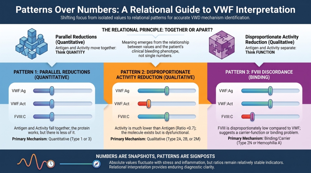
Do these values move together or apart?
If VWF antigen (VWF:Ag) and platelet-dependent VWF activity move together, think first about quantity.
If activity separates from antigen, think first about function.
If VWF collagen-binding activity (VWF:CB) is disproportionately reduced relative to VWF:Ag, think about high-molecular-weight multimer-sensitive function or selected collagen-binding defects.
If FVIII activity (FVIII:C) separates from VWF, think about carrier function or an FVIII-centered mimic.
If platelet count is low in the right context, especially with a pattern suggestive of enhanced VWF-platelet interaction, think about type 2B VWD, platelet-type VWD, or acquired VWF-platelet interaction problems.
That first-pass question often determines whether further testing is necessary and in what direction.2
The most useful comparisons in first-line VWD interpretation are summarized below.
The Comparisons That Reveal Mechanism
| Compare | Ask | Mechanistic inference |
|---|---|---|
| VWF:Ag ↔ platelet-dependent VWF activity | Do they move together or apart? | Quantity vs function |
| Platelet-dependent VWF activity ↔ multimers / VWF:CB | Is impaired function explained by architecture? | Structure–function relationship |
| VWF measurements ↔ FVIII:C | Is carrier function preserved? | VWF–FVIII binding vs FVIII-centered disorder |
| Laboratory pattern ↔ bleeding phenotype | Does the biology fit the patient? | Clinical coherence |
Table. The comparisons that reveal mechanism in VWD testing. Individual laboratory values answer different biologic questions, but diagnostic meaning emerges from the relationships among them. These comparisons transform isolated measurements into mechanistic hypotheses that guide further evaluation.
The hidden trap of single values
A VWF antigen of 42 IU/dL might reflect:
- physiologic variation or context, including ABO blood group, stress, inflammation, pregnancy, acute illness, exercise, aging, estrogen exposure, or thyroid status
- a true quantitative or clearance defect
- assay-related artifact
- multifactorial bleeding, in which VWF is contributory but not the only driver
The number itself cannot tell you which one is true. That ambiguity is built into VWF biology and into VWF measurement.3
Only pattern can.
The relational principle
Every VWF panel should be read as a system of comparisons.
Each comparison isolates a different functional dimension of VWF:
- VWF:Ag vs platelet-dependent VWF activity: quantity vs platelet-binding function
- activity vs multimer distribution: function vs structure
- VWF:CB vs VWF:Ag: collagen-binding and multimer-sensitive function vs quantity
- VWF:Ag and platelet-dependent VWF activity vs FVIII:C: VWF quantity/function vs carrier-function consequence
- VWF pattern vs platelet count: VWF behavior vs platelet clearance or platelet-type mimic
- laboratory pattern vs phenotype: signal vs lived bleeding
These comparisons turn measurements into mechanisms.4
Most VWF panels can be organized into three dominant relational patterns.
Pattern 1: parallel reductions
Quantitative pattern
If antigen and platelet-dependent activity fall together, with the activity:antigen ratio roughly preserved, the simplest inference is:
- VWF quantity is reduced
- function per molecule is relatively preserved
This pattern typically reflects:
- type 1 VWD patterns
- type 3 VWD, when VWF is absent or nearly absent
- some increased-clearance phenotypes at baseline
- physiologic low-VWF states
Key inference: the protein works, but there is less of it.5
That shorthand is useful, but it should not be overread. Type 1 VWD is heterogeneous, and increased clearance or subtle qualitative features may coexist with an apparently quantitative baseline pattern.
The therapeutic implication is conceptual rather than automatic. Therapies that depend on releasing endogenous VWF, such as desmopressin, are more likely to make sense when the released protein is functionally intact, but the response still depends on baseline level, endothelial stores, clearance behavior, subtype, and clinical context.6
Pattern 2: disproportionate activity reduction
Qualitative pattern
If platelet-dependent activity is much lower than antigen, the relationship changes:
- VWF quantity is present
- VWF function is impaired
Most diagnostic frameworks use the activity:antigen ratio as the first trigger for suspected qualitative dysfunction. An activity:antigen ratio in the approximate 0.6 to 0.7 range, depending on assay method and local validation, should prompt consideration of type 2-like mechanisms and second-line testing. The 2021 ASH/ISTH/NHF/WFH diagnostic guideline favors a cutoff of <0.7 for suspected type 2A, 2B, or 2M VWD in patients with an abnormal initial VWD screen.7
Key inference: the molecule exists, but does not behave normally.
The next mechanistic question is whether the functional loss is explained by abnormal multimer architecture, altered platelet binding, altered collagen binding, or an assay-specific problem.8
Pattern 3: FVIII discordance
Binding or carrier-function pattern
FVIII is not just another number in the panel. It is a downstream reporter of VWF’s carrier role.
If FVIII is disproportionately low compared with VWF, the pattern should raise two possibilities:
- impaired VWF-FVIII interaction, as in a type 2N mechanism
- an FVIII-centered disorder, especially mild hemophilia A
This is a carrier-function problem until proven otherwise, but the carrier may be VWF or the problem may be FVIII itself.
This is one of the clearest examples of relational interpretation. The clue is not the FVIII value alone, but the relationship between FVIII and VWF. Confirmatory approaches include the VWF-FVIII binding assay (VWF:FVIIIB) and, in selected cases, genetic testing of VWF and/or F8.9
Key inference: the problem may lie in binding, or in FVIII itself, rather than in VWF quantity.
Ratios matter because relationships matter
Absolute VWF values move.
They move with stress, inflammation, pregnancy, acute illness, aging, exercise, estrogen exposure, thyroid status, and blood type.10
Ratios do not simply summarize relationships. They often help preserve proportional relationships across changing physiologic conditions.
But ratios are still assay-dependent. They can be distorted by analytical variability, local assay performance, and the particular platelet-binding method used. A ratio is most useful when the laboratory understands its own method, reference behavior, and limitations.11
Ratios localize mechanism.
They do not eliminate ambiguity.
Why interpretation requires restraint
VWF is dynamic. A single panel is a snapshot, not a final identity.
Repeat testing is therefore not just confirmation.
It is signal clarification.
On repeat testing, the key question is not only whether a value crosses a diagnostic threshold. It is whether the relationship among values persists.
Does activity remain proportionate to antigen?
Does FVIII remain disproportionately low?
Does collagen binding remain disproportionately reduced?
Does the phenotype fit the laboratory signal?
If the panel pattern and bleeding history are discordant, the next step is to reassess both. Do not force a subtype to fit the numbers.
In VWD, persistence of pattern is often more informative than the absolute value at any single time point.12
The multimer dimension
When the panel suggests a qualitative mechanism, multimer analysis becomes structurally informative.
High-molecular-weight multimers are the most hemostatically effective forms of VWF. Loss or distortion of the multimer distribution can explain a low activity signal and can help separate qualitative subtypes. Multimer analysis is usually treated as second-line testing, triggered by an abnormal screening pattern rather than ordered reflexively for everyone.13
The structural lesson is worth making explicit:
- activity reflects function
- multimers reflect architecture
- many qualitative patterns are structure-function stories in disguise
Laboratory classification studies that link VWF sequence variants to multimerization, platelet binding, collagen binding, FVIII binding, clearance, and phenotype are especially useful because they show that subtype labels are not arbitrary categories. They are attempts to name recurring structure-function patterns.14
Collagen binding as a structural clue
VWF collagen-binding assays add another relational dimension.
Many VWF:CB assays, especially those using type I and/or type III collagen, are sensitive to high-molecular-weight multimers and can serve as a practical surrogate for multimer-sensitive function. This makes collagen binding a function that is especially sensitive to multimer size.
A reduced VWF:CB/VWF:Ag ratio may therefore point toward loss of the largest multimers, as in type 2A or 2B patterns, or toward selected, uncommon collagen-binding defects that may otherwise be missed by platelet-binding assays.15
The key point is not that collagen binding replaces multimer analysis in all settings.
The key point is that it asks a different question:
Can the VWF molecule engage collagen-dependent hemostasis in a way that matches its measured quantity?
That question belongs naturally in a relational approach.
Clearance patterns
Apparent quantitative patterns may conceal distinct mechanisms.
Some patients look “quantitative” at baseline because antigen and activity fall together, but the mechanism is not simply low production.
One tool for exploring this possibility is the VWF propeptide/VWF antigen ratio (VWFpp/VWF:Ag ratio). VWF propeptide and mature VWF are secreted together, but they have different clearance behavior. An increased VWFpp/VWF:Ag ratio can support increased clearance as a mechanism.16
Another practical probe is the desmopressin response. In some increased-clearance phenotypes, VWF may rise after desmopressin but fall more rapidly than expected, making the response short-lived.17
This finding should be interpreted modestly.
VWFpp is a mechanistic clue, not a perfect discriminator. Desmopressin response is a clinical probe, not a subtype label by itself. Both belong inside a pattern-based interpretation rather than replacing it.
When numbers mislead
Some misleading results are not clinical. They are assay-behavior problems.
A classic example is that some VWF sequence variants can affect ristocetin-dependent activity assays without necessarily reflecting impaired in-vivo hemostatic function. The best-known example is the p.D1472H variant, which can lower VWF:RCo by altering ristocetin-dependent interaction without necessarily indicating a true platelet-binding defect. This can create a false qualitative pattern on paper.18
For this reason, modern diagnostic approaches increasingly emphasize newer platelet-dependent VWF activity assays, particularly GPIb-based methods such as VWF:GPIbM and VWF:GPIbR, rather than relying solely on the older VWF:RCo assay. These assays still require careful correlation with bleeding phenotype, assay method, and local laboratory performance, because no static in-vitro assay perfectly reproduces shear-dependent VWF biology.19
Pattern-based interpretation helps because it pushes the clinician to ask:
- Is this discrepancy consistent across assays?
- Does it persist on repeat testing?
- Does it fit the multimer pattern?
- Does it fit the collagen-binding pattern?
- Does it fit the bleeding phenotype?
A ratio is a clue.
It is not a verdict.
The subtype mistake clinicians make
A common error is to assign subtype before mechanism is clear.
The wrong sequence is:
abnormal value → subtype label → therapy
The better sequence is:
pattern → mechanism → targeted second-line tests → subtype, if needed → therapy
Second-line studies exist to explain a pattern. These may include multimer analysis, VWF:CB, platelet-binding tests, RIPA in selected situations, VWF-FVIII binding assays, desmopressin response assessment, and selected genetic testing.20
Subtype is a conclusion, not a starting point.
Why relational thinking changes care
Pattern recognition:
- reduces overdiagnosis from borderline single values
- prevents premature subtype labeling
- identifies when advanced testing is necessary
- protects against assay artifacts
- anticipates which therapeutic strategies are most coherent with mechanism
Pattern recognition is not academic.
It directly shapes management.21
Clinical synthesis
The expert move in VWD testing is not to ask, “Which value is abnormal?”
It is to ask, “What relationship among values is being revealed?”
That shift changes the diagnostic task. The clinician is no longer sorting isolated numbers into boxes, but reading a biologic system: quantity, function, structure, carrier role, assay behavior, and bleeding phenotype.
In VWD, numbers matter.
But relationships explain their meaning.
Guideline perspective: relationships before subtype labels
Current diagnostic guidance does not treat VWF testing as a single-value exercise. The initial laboratory assessment is built around a panel: VWF antigen, platelet-dependent VWF activity, and FVIII activity, with additional studies such as VWF collagen binding, multimer analysis, VWF-FVIII binding, RIPA, desmopressin response, or genetic testing used selectively to explain the pattern.
The central interpretive move is relational. A preserved activity:antigen relationship points toward a quantitative pattern; a disproportionately low ratio points toward a qualitative pattern and should trigger confirmatory classification testing rather than establish a subtype by itself; and a disproportionately low FVIII level relative to VWF raises the possibility of impaired VWF-FVIII binding or an FVIII-centered mimic. Collagen binding and multimer analysis add a structure-function dimension, especially when type 2 mechanisms are suspected.
Guidelines also emphasize restraint. VWF is affected by physiologic state, inflammation, pregnancy, exercise, age, blood group, preanalytical handling, and assay method. Borderline or discordant results should therefore be interpreted using repeat testing on separate occasions when appropriate, local assay performance, preanalytical conditions, and bleeding phenotype. Ratios are useful because they reveal relationships, but they are not diagnostic verdicts.
The practical sequence is:
pattern → mechanism → targeted second-line testing → subtype if needed → therapy
Subtype assignment should be the result of interpretation, not the starting point.
Reflect & Apply
A 32-year-old woman with heavy menstrual bleeding and recurrent epistaxis has the following results:
- VWF antigen (VWF:Ag): 78 IU/dL
- Platelet-dependent VWF activity: 32 IU/dL
- FVIII activity (FVIII:C): 85 IU/dL
Most likely mechanistic category: qualitative pattern.
Platelet-dependent VWF activity is disproportionately reduced relative to VWF:Ag, with a VWF activity/VWF:Ag ratio of approximately 0.4. FVIII:C is not disproportionately reduced relative to VWF:Ag, so the presenting pattern does not suggest impaired VWF–FVIII binding or an isolated FVIII deficiency.
A parallel reduction in activity and antigen would instead suggest a quantitative pattern.
The next step is not to name the subtype immediately.
The next step is to confirm the pattern and ask what explains the activity-antigen mismatch:
- loss of high-molecular-weight multimers?
- altered platelet-binding function?
- does VWF:CB show a concordant multimer-sensitive abnormality?
- assay artifact?
- a type 2-like qualitative mechanism requiring second-line testing?
This pattern should trigger repeat confirmation when appropriate and targeted second-line testing before subtype labeling.
The pattern points toward mechanism.
The mechanism guides the next test.
Test your thinking
A short quiz on interpreting VWF labs relationally in VWD.