
Why diagnosing von Willebrand disease is a reasoning problem, not a test result
Note: The video and audio linked above were generated with the assistance of AI. Clinical accuracy has been reviewed, but no AI-generated content can be guaranteed to be fully error-free.
Why this spoke matters
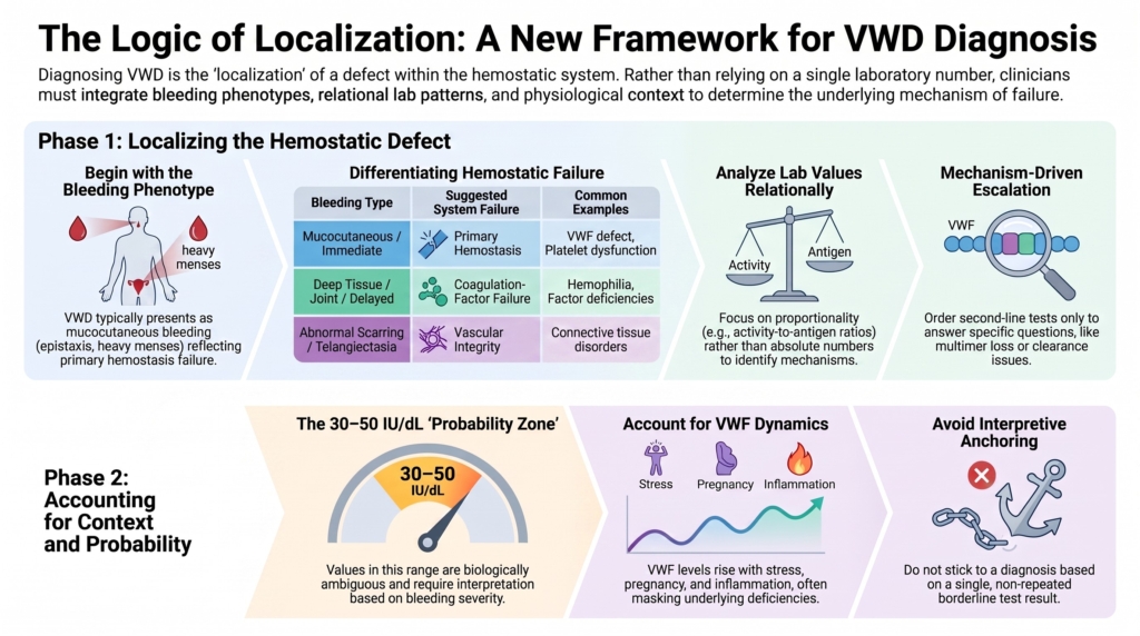
Diagnosing von Willebrand disease (VWD) is often framed as ordering the correct laboratory panel.
That framing is understandable.
Guidelines list recommended assays, cutoffs, VWF activity/VWF:Ag ratios, and subtype-directed tests.1
But diagnosis is not the detection of a number.
It is the localization of a defect within the hemostatic system.
By localization, we mean identifying which component of hemostasis is failing and, when possible, which function within that component is impaired.
That process has two modes.
Sometimes the task is true localization: the pattern points to a discrete mechanism, such as loss of high-molecular-weight multimers, increased platelet binding, defective FVIII binding, or accelerated VWF clearance.
At other times, localization reaches its limit. The phenotype is mild, the VWF value is borderline, the signal is context-sensitive, and the task becomes probability estimation rather than lesion-finding.
Expert diagnosis requires knowing which mode one is in.
The ASH/ISTH/NHF/WFH diagnostic guideline structures evaluation around bleeding phenotype, VWF antigen (VWF:Ag), platelet-dependent VWF activity, factor VIII activity (FVIII:C), VWF activity/VWF:Ag relationships, subtype testing, and reassessment when levels normalize over time.2
Tosetto and Eikenboom make the probabilistic logic explicit: the same laboratory value has different diagnostic meaning depending on the clinical setting, pre-test probability, bleeding phenotype, and family context.3
Laboratory values inform inference.
They do not constitute diagnosis.
Diagnosis begins with phenotype
VWD is usually suspected because the bleeding pattern suggests a problem in primary hemostasis.
Typical symptoms include:
- mucocutaneous bleeding
- epistaxis
- easy bruising
- heavy menstrual bleeding
- oral cavity bleeding
- prolonged bleeding after dental work, childbirth, surgery, or minor wounds
This pattern reflects VWF biology. VWF supports platelet adhesion under shear and stabilizes circulating factor VIII.4
Deep muscle bleeding, hemarthrosis, or large soft-tissue hematomas point more strongly toward a coagulation-factor mechanism unless VWF deficiency is severe, as in type 3 VWD or type 2N VWD with markedly reduced FVIII.5
Validated bleeding assessment tools help standardize the history. They are especially useful in low-prevalence settings to decide who should undergo VWF-specific testing. In referred or high-probability settings, they should not be used as the sole reason to withhold testing.6
A bleeding score is not a verdict.
It is a structured way of asking whether the phenotype is plausible.
Step 1: Localize the bleeding system
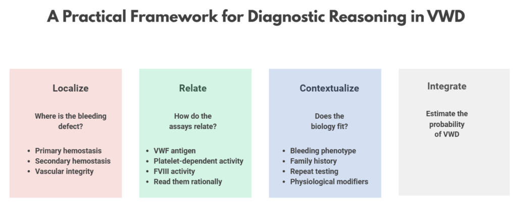
The first diagnostic question is not “What is the VWF level?”
It is:
Which hemostatic subsystem seems abnormal?
The main possibilities are:
- primary hemostasis: platelet number, platelet function, VWF, or vessel-wall interaction
- secondary hemostasis: coagulation-factor generation and fibrin formation
- vascular integrity: connective tissue, vascular malformations, or structural vessel fragility
These categories overlap in practice, but they provide a useful first approximation.
A practical heuristic:
- mucocutaneous, immediate bleeding suggests primary hemostasis
- deep tissue, joint, or delayed bleeding suggests coagulation-factor failure
- bleeding with hypermobility, abnormal scarring, telangiectasias, or vessel fragility suggests vascular or connective-tissue mechanisms
VWF sits at an interface. It helps platelets adhere where shear is high, and it protects FVIII from premature clearance.7
That dual role explains why most VWD behaves like a primary hemostatic disorder, while severe VWF deficiency or type 2N VWD can resemble a coagulation-factor disorder.
This upstream triage matters.
A borderline VWF value in a patient with heavy menstrual bleeding and epistaxis is not the same as the same value in a patient with isolated hemarthroses.
The number is interpreted through the phenotype.
Step 2: Ask whether VWF is plausibly involved
If the bleeding phenotype fits primary hemostasis, VWF testing becomes appropriate.
First-level VWD testing usually includes:
- VWF antigen (VWF:Ag)
- platelet-dependent VWF activity
- FVIII activity (FVIII:C)
A complete blood count, platelet count, PT, aPTT, and fibrinogen help evaluate other bleeding mechanisms, but normal PT and aPTT do not exclude VWD.8
The laboratory pattern should be read relationally:
- parallel reduction of VWF:Ag and platelet-dependent VWF activity suggests a quantitative deficiency pattern
- disproportionately reduced platelet-dependent VWF activity relative to VWF:Ag suggests a qualitative VWF defect
- disproportionately reduced FVIII:C relative to VWF:Ag suggests type 2N VWD or hemophilia A
The key is proportionality, not the absolute value, because proportionality encodes mechanism.
A VWF activity/VWF:Ag ratio below approximately 0.7 should raise concern for type 2 VWD. and prompt a subtype-directed question, though the exact cutoff and interpretation depend on assay method and local validation.9
Modern VWF glycoprotein IbM activity (VWF:GPIbM) and VWF glycoprotein IbR activity (VWF:GPIbR) assays are generally preferred over classic VWF ristocetin cofactor activity (VWF:RCo) testing when available, because ristocetin cofactor testing is less precise, performs poorly at low levels, and can be misleading in the presence of ristocetin-sensitive polymorphisms that do not reflect true VWF dysfunction.10
This is the transition from:
Is VWF low?
to:
Does the pattern make mechanistic sense?
Step 3: Escalate only when there is a mechanistic question
Second-line testing should not be reflexive after any single abnormal value.
It should be driven by a specific question.
If platelet-dependent activity is disproportionately low, the question becomes whether high-molecular-weight multimers are missing or whether ligand binding is intrinsically defective.
If FVIII:C is disproportionately low, the question becomes whether VWF is failing to bind FVIII.
If platelet count is low or type 2B is suspected, the question becomes whether VWF is binding platelet glycoprotein Ib (GPIb) too avidly.
If DDAVP produces a brisk early rise followed by rapid decline, the question becomes whether accelerated clearance is present.
The relevant tests include VWF multimer analysis, VWF collagen-binding activity (VWF:CB), RIPA or targeted genetic testing for type 2B, VWF–FVIII binding assay (VWF:FVIIIB) or genetic testing for type 2N, and desmopressin response kinetics or VWF propeptide (VWFpp) testing for clearance phenotypes.11
Subtype classification is therefore a summary of mechanism.
It is not a memorized label attached to an isolated number.
Step 4: Ask whether the signal is intrinsic or contextual
VWF is dynamic.
Levels can rise with stress, inflammation, exercise, pregnancy, estrogen exposure, acute bleeding, comorbid illness, and aging.12
Preanalytical issues also matter. Sample handling, refrigeration, delayed processing, transport conditions, freezing and thawing, and assay imprecision can create false-normal or false-abnormal patterns.13
Failure to account for context is a major source of error in mild or borderline phenotypes.
Testing should generally be repeated under baseline conditions before a durable label is assigned, especially when results are borderline, discordant, or biologically implausible.14
Sometimes the most important next test is not an expanded panel.
It is repeating the first-line tests (VWF:Ag, platelet-dependent VWF activity, and FVIII:C correctly) correctly.
Step 5: Integrate inheritance and genetics without overreading them
Classic diagnostic reasoning includes:15
- personal bleeding history
- family history
- reduced or dysfunctional VWF
- inheritance pattern
- subtype-consistent laboratory phenotype
Type 1 and many type 2 variants are often autosomal dominant. Type 3 and type 2N are usually autosomal recessive. Type 1 VWD may show incomplete penetrance and variable expressivity, and mild quantitative phenotypes may reflect modifiers beyond the VWF gene.16
Genetic testing is useful when it answers a specific diagnostic question:
- Is this type 2B VWD or platelet-type VWD?
- Is this type 2N VWD or mild hemophilia A?
- Is this type 3 VWD relevant for family counseling?
- Is this ambiguous type 2 pattern genetically supported?
It is less useful as a routine diagnostic solution for most type 1 or borderline quantitative phenotypes, where variants may be absent, polygenic, or of uncertain significance.17
Genotype can support localization.
It should not erase phenotype.
Step 6: Know when localization becomes probability estimation
Some VWD diagnoses are mechanistically clear.
Others are not.
This is most evident in the 30-50 IU/dL range, where VWF:Ag or platelet-dependent VWF activity may be mildly reduced, bleeding symptoms may be present, and the causal contribution of VWF may be uncertain.
The 2021 ASH/ISTH/NHF/WFH guideline recommends diagnosing type 1 VWD when VWF:Ag and/or platelet-dependent VWF activity is below 30 IU/dL regardless of bleeding, and when VWF:Ag and/or platelet-dependent VWF activity is 30–50 IU/dL in the presence of abnormal bleeding.18
The BSH laboratory guideline emphasizes caution in the 30-50 IU/dL range, noting that reduced VWF in this range may be insufficient by itself to explain bleeding and that additional hemostatic abnormalities may contribute.19
Both perspectives matter.
One protects access to care for patients with real bleeding, especially those with heavy menstrual bleeding and procedure-related bleeding.
The other protects patients from unnecessary medicalization and premature diagnostic closure.
The practical question is not:
Does this patient cross a cutoff?
It is:
Does the total evidence justify a disease label and its consequences?
Why misdiagnosis happens
VWD is vulnerable to both underdiagnosis and overdiagnosis.
Underdiagnosis happens when clinicians normalize heavy menstrual bleeding, dismiss recurrent mucocutaneous bleeding, fail to ask about procedure-related bleeding, or assume that normal screening coagulation tests exclude VWD.
Overdiagnosis happens when clinicians anchor on a single borderline VWF value, ignore phenotype, fail to repeat testing, or mistake low VWF as the only possible explanation for bleeding.20
The errors fall into three broad groups:
- Testing errors: sampling during stress, pregnancy, inflammation, acute bleeding, exercise, or poor sample handling
- Interpretive errors: treating platelet-dependent VWF activity, a VWF activity/VWF:Ag ratio, or a borderline VWF value as definitive
- Anchoring errors: keeping an old diagnosis without reassessment or stopping the workup once a mildly low VWF level is found
The conceptual error is the same:
mistaking a measurement for the mechanism that produced it.
The acquired-mimic problem
Not every VWF abnormality is inherited VWD.
Acquired von Willebrand syndrome can produce laboratory patterns that resemble congenital VWD, including loss of high-molecular-weight multimers in high-shear states.21
This matters because localization must include the question:
Is the VWF abnormality inherited, acquired, or contextual?
A type 2A-like pattern in a child with lifelong bleeding and family history means something different from a similar pattern in an older adult with new bleeding and a high-shear cardiac lesion.
Same pattern.
Different diagnosis.
Different treatment.
Clinical consequences of correct localization
Mechanism determines management.
A quantitative deficiency pattern may predict DDAVP responsiveness, especially in many patients with type 1 VWD.
A clearance phenotype may show a brisk early DDAVP response but a short-lived effect.
Type 2B requires caution because DDAVP may worsen thrombocytopenia.
Type 2N requires recognition that FVIII:C is low because VWF is failing to protect it, not because the patient necessarily has hemophilia A.
Type 3 generally requires VWF replacement.22
Treatment is not chosen by label alone.
It is chosen by mechanism, bleeding phenotype, procedure risk, and expected duration of hemostatic challenge.
A patient may not meet strict criteria for definitive VWD and still require thoughtful hemostatic planning for childbirth, dental extraction, surgery, or mucosal procedures.
Diagnosis and treatment planning overlap.
They are not identical.
A practical bedside heuristic
Ask three questions:
- Does the bleeding phenotype fit VWF biology?
- Does the laboratory pattern reflect a coherent mechanism?
- Does the probability justify a disease label?
If the answer to any question is no, pause.
Repeat testing if needed.
Re-localize the bleeding.
Consider platelet disorders, connective tissue disorders, gynecologic causes, vascular lesions, acquired VWF abnormalities, and other rare bleeding disorders.
The goal is not to prove VWD.
The goal is to understand the bleeding.
Evidence anchor: why VWD diagnosis requires localization and probability
Summary derived from diagnostic guidelines, laboratory standards, cohort studies, and expert reviews. The evidence consistently supports a diagnostic model that integrates bleeding phenotype, first-line VWF testing, assay relationships, physiologic context, inheritance, and selective second-line testing. No single VWF value, bleeding score, family-history pattern, or genetic result is sufficient in isolation.
| Evidence stream | What it shows | Why it matters | Main limitation |
|---|---|---|---|
| Bleeding phenotype and BATs | VWD typically produces mucocutaneous bleeding, including epistaxis, easy bruising, heavy menstrual bleeding, oral bleeding, and procedure-related bleeding. BATs help standardize history and are most useful in low-prevalence settings. | Diagnosis should begin with phenotype and pre-test probability, not reflex laboratory testing. | BATs may be falsely reassuring in children or patients without hemostatic challenges, and positive scores are not specific for VWD. |
| First-line VWF testing | VWF antigen (VWF:Ag), platelet-dependent VWF activity, and factor VIII activity (FVIII:C) define the initial laboratory pattern. | The key interpretive move is relational: VWF:Ag and platelet-dependent VWF activity falling together suggests quantitative deficiency; disproportionate activity reduction suggests qualitative VWF dysfunction; disproportionate FVIII:C reduction raises type 2N VWD or hemophilia A. | Assays vary by method, laboratory, sample handling, and biologic context. A single result may mislead. |
| VWF activity/VWF:Ag ratios | A low platelet-dependent VWF activity/VWF antigen ratio, often using a cutoff near 0.7, supports concern for type 2 VWD and prompts subtype-directed evaluation. | Ratios help convert isolated values into mechanistic patterns. | Cutoffs vary across guidelines and assay systems; ratios are screening signals, not final subtype diagnoses. |
| Second-line testing | VWF multimer analysis, VWF collagen-binding activity (VWF:CB), RIPA or targeted genetic testing, VWF–FVIII binding assay (VWF:FVIIIB), desmopressin response kinetics, and VWF propeptide (VWFpp) testing refine the mechanism. | Second-line testing should answer a specific localization question, not function as a reflex “bigger panel.” | Some tests are technically demanding, variably available, and difficult to interpret without expertise. |
| VWF dynamics and repeat testing | VWF levels vary with stress, inflammation, pregnancy, exercise, estrogen exposure, aging, acute bleeding, comorbid illness, and preanalytical handling. | Borderline or discordant results should usually be repeated under baseline conditions before assigning a durable label. | Repeat testing may still leave uncertainty, especially in mild quantitative phenotypes. |
| Low VWF and threshold uncertainty | Values in the 30–50 IU/dL range are clinically and biologically ambiguous. Current guidelines differ in terminology and emphasis, especially around “low VWF” versus type 1 VWD with bleeding. | This range requires probability estimation, not automatic labeling or dismissal. | Thresholds are useful for care access and standardization but do not define a sharp biologic boundary. |
| Genetic testing | Genetic testing is most useful for selected type 2 variants, type 2B versus platelet-type VWD, type 2N versus hemophilia A, and type 3 family counseling. | Genotype can support localization when the question is specific. | Genetic testing is often less helpful in type 1 or borderline quantitative phenotypes, where variants may be absent, polygenic, or of uncertain significance. |
Interpretive note: VWD diagnosis is strongest when the bleeding phenotype, laboratory pattern, inheritance context, and physiologic setting converge on a coherent mechanism. When they do not converge, the correct response is not to force the label, but to re-localize the bleeding problem.
Guideline perspective: diagnostic localization in VWD
Based on international diagnostic guidelines, laboratory standards, and expert reviews.
Guideline organizations and sources referenced
- ASH/ISTH/NHF/WFH diagnostic guideline
- BSH/UKHCDO laboratory diagnosis guideline
- NHLBI diagnostic framework
- Expert reviews on VWD diagnosis, low VWF, assay interpretation, and “undiagnosing” VWD
Shared guidance themes
- Start with bleeding phenotype and pre-test probability.
- Use VWF antigen (VWF:Ag), platelet-dependent VWF activity, and factor VIII activity (FVIII:C) as first-line tests.
- Interpret VWF testing relationally, especially VWF activity/VWF:Ag and FVIII:C/VWF:Ag patterns.
- Repeat borderline or discordant testing under baseline conditions.
- Use second-line testing only when it answers a specific mechanistic question.
- Treat VWF:Ag or platelet-dependent VWF activity in the 30–50 IU/dL range as context-dependent rather than automatically normal or automatically diagnostic.
- Reassess historical VWD diagnoses when current phenotype and laboratory data no longer support the label.
What guidelines emphasize
- VWD cannot be diagnosed or excluded by PT/aPTT alone.
- BATs help standardize bleeding history but do not replace clinical judgment.
- Assay method and preanalytical handling matter.
- Subtype classification is clinically important because mechanism affects treatment.
What guidelines do not support
- Diagnosing VWD from a single borderline VWF value.
- Using blood group O to dismiss a convincing bleeding phenotype.
- Ordering second-line tests without a defined diagnostic question.
- Treating genetic testing as a substitute for phenotype and VWF laboratory interpretation.
- Assuming normalized VWF levels with age automatically erase bleeding risk.
Reflect & Apply Case
A patient has:
- recurrent delayed deep muscle hematomas after minor trauma
- no epistaxis
- no heavy menstrual bleeding
- no dental bleeding
- normal platelet count
- normal PT and aPTT
- VWF:Ag 43 IU/dL
- Platelet-dependent VWF activity: 41 IU/dL
- FVIII:C 96 IU/dL
- blood group O
- no family history of mucocutaneous bleeding
What is the most justified interpretation?
VWF:Ag and platelet-dependent VWF activity are proportionally reduced, which argues against a qualitative type 2 pattern. The value lies in the 30-50 IU/dL range, where the meaning depends heavily on phenotype, testing conditions, and repeat measurement.
But the bleeding phenotype does not strongly localize to VWF biology. Delayed deep muscle hematomas point away from classic primary hemostatic dysfunction.
Best interpretation:
Mild proportional VWF reduction, possibly low VWF or a type 1-range quantitative signal, but the bleeding phenotype is not well explained by VWF alone. Repeat VWF testing under baseline conditions is reasonable, but the diagnostic priority is to re-localize the bleeding mechanism rather than assign VWD from the number.
A lower VWF value, a strong mucocutaneous phenotype, or a convincing family history would shift the probability toward VWD.
Here, the pattern asks for caution.
Not dismissal.
Not a premature label.
Localization first.
Test your thinking
A short quiz on diagnostic approach in VWD.