
When patterns demand deeper localization
Note: The video and audio linked above were generated with the assistance of AI. Clinical accuracy has been reviewed, but no AI-generated content can be guaranteed to be fully error-free.
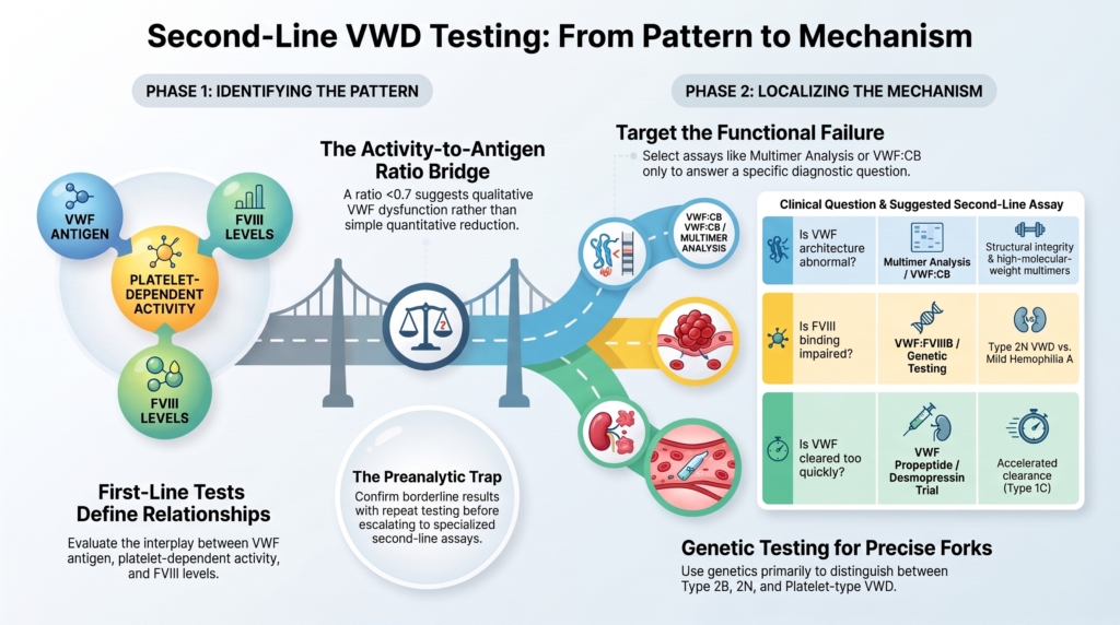
Why this spoke matters
Most patients evaluated for von Willebrand disease do not need every VWF-related assay.
They need a coherent diagnostic sequence.
First-line tests ask:
- is VWF reduced?
- is platelet-dependent VWF activity reduced?
- is FVIII reduced?
- do these values relate to one another in a recognizable pattern?
Second-line tests ask a different question:
Where, within the VWF system, is the failure likely to be?
That distinction matters.
Second-line testing is not “screening harder.”
It is characterization.
It is localization.
It is the next step only when the first-line pattern has created a specific mechanistic question. Contemporary diagnostic approaches begin with bleeding phenotype and first-level laboratory testing, usually including VWF antigen (VWF:Ag), platelet-dependent VWF activity, and factor VIII activity (FVIII:C). In this essay, “platelet-dependent VWF activity” refers to assays such as VWF:GPIbM, VWF:GPIbR, or VWF:RCo, depending on the method used by the laboratory. Second-level tests are then used to characterize type 2 variants, increased clearance, VWF–FVIII binding defects, and other specific mechanisms.1
A patient with an otherwise typical mild type 1 or low VWF pattern, with VWF:Ag and platelet-dependent VWF activity moving together, does not automatically need multimers, RIPA, VWF–FVIII binding assay (VWF:FVIIIB), VWF propeptide (VWFpp) testing.
A patient with disproportionate loss of activity, unexplained thrombocytopenia, unusually low FVIII, abnormal collagen binding, or a short-lived desmopressin response may.
The principle is simple:
Do not order a second-line test until you can say what question it is supposed to answer.
From pattern to mechanism
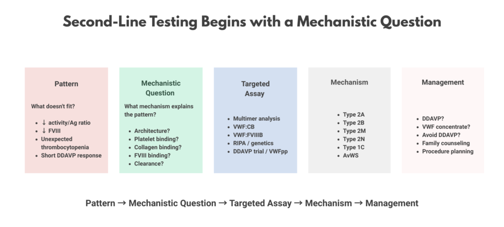
A useful sequence is:
- bleeding phenotype
- first-line VWF pattern
- mechanistic question
- targeted second-line assay
- subtype interpretation, if needed
- therapeutic implication
This is what diagnostic reasoning as localization means.
The clinician is not simply trying to prove that VWD is present.
The clinician is asking:
- is this primarily quantitative or qualitative?
- is VWF–platelet binding reduced or increased?
- is collagen binding abnormal?
- is multimer structure abnormal?
- is VWF–FVIII binding impaired?
- is VWF being cleared too quickly?
- is this inherited VWD, platelet-type VWD, mild hemophilia A, acquired von Willebrand syndrome, or something else?
Second-line tests are useful when they narrow one of those forks.
They are less useful when the diagnostic problem is not yet well framed.
First-line tests identify relationships
Before second-line testing, the clinician needs to understand the relationship among the first-line values.
The usual first-line VWD pattern includes:
- VWF antigen (VWF:Ag)
- platelet-dependent VWF activity
- FVIII activity (FVIII:C)
- CBC and platelet count
- PT and aPTT where appropriate
- bleeding history and family history
These tests do not simply generate numbers.
They define relationships.
A proportional reduction in VWF:Ag and platelet-dependent VWF activity suggests one kind of problem.
A disproportionate reduction in platelet-dependent activity relative to antigen suggests another.
A disproportionately low FVIII:C level raises a different question.
A low platelet count with abnormal platelet-dependent VWF activity changes the map again.
A normal aPTT does not exclude VWD, and a platelet function analyzer should not be used as a stand-alone screen to rule out VWD when clinical suspicion is real.2
Second-line testing begins when these relationships point somewhere.
If the pattern is borderline, unstable, or biologically implausible, repeat first-line testing under better conditions before ordering specialized assays.
VWF activity/VWF:Ag ratio: the bridge to characterization
The VWF activity/VWF:Ag ratio is not a second-line assay in itself.
It is the bridge between first-line testing and second-line characterization.
A low VWF activity/VWF:Ag ratio suggests that the VWF protein present in plasma is not functioning proportionally.
The amount may not be the whole problem.
The molecule may be present but qualitatively impaired.
The 2021 ASH/ISTH/NHF/WFH diagnostic guideline suggests using a VWF activity/VWF:Ag ratio cutoff of <0.7, rather than <0.5, when type 2A, 2B, or 2M VWD is suspected in the setting of an abnormal initial VWD screen.3
But ratios should not be treated as magic.
They are directional clues.
They ask:
Is there a qualitative defect in VWF function?
The answer may depend on:
- multimer analysis
- collagen-binding testing
- platelet-binding studies
- the specific activity assay used
- repeat testing before escalation
Ratios do not name the subtype by themselves.
They tell the clinician where to look next.
Assay choice matters
Historically, VWF ristocetin cofactor activity, or VWF:RCo, was the standard platelet-dependent activity assay.
It remains important, but it is also imperfect.
VWF:RCo uses ristocetin, a nonphysiologic antibiotic, to induce VWF binding to platelet GPIb. The assay has limitations, including variability, reduced sensitivity at low VWF levels, and falsely low results in some individuals with variants that affect ristocetin binding rather than physiologic VWF function.4
Newer assays, including VWF glycoprotein IbM activity (VWF:GPIbM) and VWF glycoprotein IbR activity (VWF:GPIbR), assess platelet-binding activity using recombinant GPIb-based methods. The 2021 diagnostic guideline suggests newer GPIb-based platelet-dependent VWF activity assays over VWF:RCo when available, although no assay is fully physiologic and local laboratory expertise remains essential.5
This matters because second-line testing is only as good as the pattern that triggers it.
If the first-line activity result is misleading, the entire diagnostic branch may point in the wrong direction.
Multimer analysis: when architecture may explain dysfunction
Multimer analysis is most useful when the first-line pattern suggests a qualitative defect.
The classic clue is a disproportionately low platelet-dependent VWF activity compared with VWF:Ag.
That pattern suggests that VWF quantity is not the whole problem.
The molecule may be present, but structurally abnormal.
Multimer analysis asks:
Is VWF architecture abnormal?
VWF circulates as multimers of varying size.
The larger multimers are especially important for platelet adhesion under shear.
When high-molecular-weight multimers are reduced or absent, platelet-dependent activity may fall out of proportion to antigen.
That pattern can support type 2A or type 2B VWD, depending on the broader laboratory and clinical context. Preserved multimers in the setting of reduced platelet-dependent activity may instead support type 2M VWD.6
Multimer analysis can help identify:
- loss of high-molecular-weight multimers
- abnormal multimer distribution
- preserved multimers despite reduced activity
- patterns that support or weaken specific subtype hypotheses
But multimer analysis does not tell the clinician how much the patient bleeds.
It does not replace the bleeding history.
It does not convert a borderline phenotype into certainty.
It localizes one possible failure mode:
VWF architecture.
VWF collagen binding: when vessel-wall adhesion is the question
VWF does not only bind platelets.
It also binds collagen exposed at sites of vascular injury.
The VWF collagen-binding assay, or VWF:CB, asks:
Can VWF bind collagen appropriately?
This matters in two key ways.
First, collagen binding is influenced by multimer size. High-molecular-weight multimers bind collagen more effectively, so VWF:CB, especially when expressed as the VWF:CB/VWF:Ag ratio, can serve as a functional clue to loss of high-molecular-weight multimers, depending on assay design and collagen source.7
Second, collagen-binding defects can occur even when the platelet-binding axis is not the dominant abnormality.
This is why VWF:CB is not merely a poor person’s multimer analysis.
It asks a related but distinct question.
The 2021 diagnostic guideline suggests either VWF multimer analysis or VWF:CB/VWF:Ag when additional testing is needed for suspected type 2A, 2B, or 2M VWD.8
Conceptually:
- low VWF activity/VWF:Ag ratio plus low VWF:CB/VWF:Ag ratio supports loss of high-molecular-weight multimers
- low platelet-dependent VWF activity with preserved VWF:CB may support a platelet-binding type 2M pattern
- isolated or disproportionate collagen-binding impairment raises the possibility of a collagen-binding defect
But VWF:CB also has limits.
Assays differ by collagen type, method, optimization, and availability. Collagen types I and III are commonly used and may be useful surrogates for high-molecular-weight multimers; other collagen-binding biology is less routinely assessed in clinical practice.9
The test is powerful when optimized.
It is misleading when treated as generic.
RIPA and targeted genetic testing: when VWF–platelet binding is increased
Ristocetin-induced platelet agglutination, or RIPA, belongs in the diagnostic pathway only when the VWF–platelet interaction is the question.
Its most important role is in suspected enhanced platelet binding, especially when type 2B VWD or platelet-type VWD is being considered.
The clinical-laboratory pattern may include:
- disproportionately low VWF activity
- loss of high-molecular-weight multimers
- thrombocytopenia, especially if intermittent or stress-related
- family pattern suggesting qualitative VWF dysfunction
- concern for type 2B VWD or platelet-type VWD
Low-dose RIPA asks:
Is platelet agglutination occurring at lower ristocetin concentrations than expected?
In normal individuals, low concentrations of ristocetin do not usually produce platelet agglutination.
In type 2B VWD, VWF has increased affinity for platelet GPIb.
In platelet-type VWD, the platelet GPIb receptor has increased affinity for VWF.
The laboratory phenotype can look similar.
The biology is different.
In type 2B VWD, the problem is usually in VWF.
In platelet-type VWD, the problem is usually in the platelet receptor.
That difference matters for family counseling, interpretation of thrombocytopenia, and treatment planning.10
RIPA is therefore not just a laboratory detail.
It is a localization test.
But the modern framing needs care.
The 2021 ASH/ISTH/NHF/WFH diagnostic guideline suggests targeted genetic testing over low-dose RIPA for suspected type 2B VWD when additional testing is needed and genetic testing is available. RIPA remains relevant mechanistically and operationally, especially where genetic testing is unavailable, delayed, or where platelet-type VWD remains in the differential.11
So RIPA should be taught in two ways.
Historically and mechanistically, it is central.
Operationally, it is one tool among several, increasingly paired with or superseded by targeted genetic testing in specialized diagnostic pathways.
VWF-FVIII binding evaluation: when FVIII:C is disproportionately low
VWF stabilizes FVIII in circulation.
That means VWD can sometimes present as a FVIII problem.
When FVIII:C is much lower than expected for the VWF:Ag and platelet-dependent VWF activity levels, the clinician should pause.
This pattern asks:
Is the problem VWF-mediated FVIII carriage, or is this primary FVIII deficiency?
This is the classic fork between type 2N VWD and mild hemophilia A.
The distinction is not semantic.
It changes the diagnosis.
It changes inheritance counseling.
It changes treatment.
It changes how the family history is interpreted.
Type 2N VWD results from impaired binding of VWF to FVIII. Patients may have low FVIII with VWF antigen and platelet-dependent activity that are not proportionally reduced, creating a phenotype that can mimic mild hemophilia A.12
VWF–FVIII binding assays (VWF:FVIIIB), and in some settings targeted genetic testing, help determine whether VWF is failing to bind FVIII appropriately.
The 2021 diagnostic guideline suggests either VWF:FVIIIB testing or targeted genetic testing for suspected type 2N VWD when additional testing is needed. The BSH laboratory guideline also supports genetic testing and consideration of VWF:FVIIIB testing in this diagnostic fork.13
This is second-line testing at its best.
The first-line pattern creates a precise question.
The second-line test answers that question.
VWF propeptide (VWFpp) and desmopressin response kinetics: when clearance is suspected
Some patients appear to have a quantitative VWF deficiency, but the mechanism is not low production alone.
The problem may be accelerated clearance.
This becomes especially relevant when:
- VWF levels are repeatedly low
- VWF:Ag and platelet-dependent VWF activity rise after desmopressin but fall rapidly
- the clinical pattern suggests short-lived correction
- the question is whether the patient has type 1C VWD or another increased-clearance phenotype
VWF propeptide (VWFpp) testing helps here because VWFpp and mature VWF are secreted together but cleared at different rates. An increased VWFpp/VWF:Ag ratio can support accelerated VWF clearance.14
The question is:
Is the low VWF signal due, at least in part, to shortened VWF survival?
This matters because a patient with accelerated clearance may have a brisk initial response to desmopressin but inadequate duration of effect.
The 2021 diagnostic guideline suggests using a desmopressin trial with 1-hour and 4-hour post-infusion testing, rather than relying on VWFpp/VWF:Ag alone, when type 1C VWD is suspected.15
The goal is not to generate another number.
It is to explain the pattern.
Genetic testing: when plasma does not settle the mechanism
Genetic testing is powerful.
But it is not always clarifying.
In VWD, its value depends heavily on the question being asked.
It is most useful when a genetic result may resolve a specific mechanistic fork, such as:
- suspected type 2B VWD
- suspected platelet-type VWD
- suspected type 2N VWD
- severe VWF deficiency where type 3 VWD is possible
- family counseling when the causative variant is known
- discordant subtype patterns where phenotypic assays are unavailable or difficult to interpret
It is often less helpful when the clinical question is mild quantitative VWF reduction, especially in the low VWF range.
The VWF gene is highly polymorphic, and many variants are not clearly pathogenic. Type 1 VWD often has incomplete genotype-phenotype correlation, whereas type 2 and type 3 VWD are more likely to have identifiable and interpretable VWF variants.16
The absence of a clearly pathogenic VWF variant does not necessarily prove that the bleeding phenotype is irrelevant.
The presence of a variant does not automatically define the patient’s future bleeding risk.
Phenotype remains the basis of classification.
Genotype supports, clarifies, or challenges that classification.
Genetics can clarify mechanism.
It does not replace clinical judgment.
The preanalytic trap
Before reaching for second-line testing, the clinician should ask whether the first-line pattern is trustworthy.
VWF levels are biologically and analytically sensitive.
They may be affected by inflammation, pregnancy, exercise, stress, acute bleeding, aging, and sample handling.
The BSH laboratory guideline emphasizes that diagnostic samples should be collected when the patient is at baseline health, that diagnosis should be based on samples collected on two separate occasions, and that preanalytic handling matters: whole blood should not be refrigerated, samples should be transported at ambient temperature, and citrated plasma for VWD assays should be tested or frozen within recommended timeframes.17
Second-line testing cannot rescue a poorly defined or poorly collected first-line pattern.
Sometimes the best “second-line” move is not a specialized assay.
It is repeating the right first-line tests under better conditions.
The acquired VWS reminder
Not every abnormal VWF pattern is inherited VWD.
Acquired von Willebrand syndrome can produce laboratory patterns that resemble inherited qualitative VWD, especially when high-molecular-weight multimers are lost.
This matters when the patient is older, has no lifelong bleeding history, has no suggestive family history, or has comorbidities associated with acquired VWF dysfunction.
The diagnostic question then changes.
It is not only:
Which VWD subtype is this?
It is also:
Is this inherited VWD at all?
Second-line results must therefore be interpreted against age of onset, family history, comorbid illness, and the broader clinical context.18
The over-testing trap
The common error is:
abnormal value → order everything
The better approach is:
pattern → question → targeted test
Ordering multiple second-line tests without a clear diagnostic question can create problems:
- incidental or ambiguous findings
- discordant results
- excessive confidence in weak signals
- delayed interpretation
- increased cost
- confusion for patients and clinicians
More testing does not always create more clarity.
Sometimes it creates a larger pile of poorly integrated data.
In VWD, this matters because the disorder lives near several boundaries:
- normal variation and disease
- type 1 VWD and low VWF
- type 2A and type 2B
- type 2A and type 2M
- type 2B and platelet-type VWD
- type 2N and mild hemophilia A
- inherited VWD and acquired von Willebrand syndrome
Second-line testing should sharpen these boundaries when possible.
It should not blur them further.
A simple localization map
If the first-line pattern suggests proportional quantitative deficiency:
- repeat testing may matter more than immediate advanced testing
- consider baseline health, inflammation, pregnancy, exercise, stress, age, hormones, and sample handling
- consider type 1C if the response to desmopressin is short-lived
- consider VWF propeptide or desmopressin response testing when increased clearance is suspected
- consider genetics selectively if it will change interpretation, counseling, or management
If the first-line pattern suggests disproportionate loss of platelet-dependent activity:
- review the activity assay used
- consider repeat confirmation
- consider VWF activity/VWF:Ag ratio
- consider multimer analysis
- consider VWF:CB/VWF:Ag ratio
- consider platelet-binding studies or genetic testing if type 2B or platelet-type VWD is plausible
If the pattern suggests loss of high-molecular-weight multimers:
- consider type 2A VWD
- consider type 2B VWD
- consider acquired von Willebrand syndrome in the appropriate clinical setting
- use RIPA and/or targeted genetic testing when enhanced platelet binding is suspected
If the pattern suggests preserved multimers with low platelet-dependent activity:
- consider type 2M VWD
- consider assay-specific limitations
- consider whether collagen binding is preserved or abnormal
If FVIII:C is disproportionately low:
- consider type 2N VWD
- consider mild hemophilia A
- use VWF–FVIII binding assay (VWF:FVIIIB) and/or targeted genetic testing
If the pattern is unstable or borderline:
- repeat testing may be the most important next step
- do not use advanced assays to force certainty prematurely
What second-line testing cannot fix
Second-line testing is powerful.
But it cannot:
- substitute for bleeding history
- determine whether symptoms are clinically meaningful by itself
- erase assay variability
- convert continuous biology into clean categories
- eliminate the need for judgment
- decide, by itself, whether treatment is needed
This is especially important in borderline disease.
A second-line test may explain a mechanism.
It may support or confirm a subtype.
It may clarify a treatment choice.
But it does not establish the whole diagnosis in isolation.
Clinical synthesis
Second-line VWD testing is best understood as boundary adjudication.
The clinician is often standing between adjacent possibilities:
- type 1 VWD or low VWF?
- type 2A or type 2M?
- type 2A or type 2B?
- type 2B or platelet-type VWD?
- type 2N or mild hemophilia A?
- inherited VWD or acquired von Willebrand syndrome?
- true VWF dysfunction or assay artifact?
Second-line tests help decide which side of the boundary the patient is likely to be on.
They do this by localizing mechanism:
- architecture
- platelet binding
- collagen binding
- FVIII binding
- clearance
- genotype
The goal is not to order more.
The goal is to ask better.
Second-line tests characterize.
They may confirm a subtype or mechanism.
They do not confirm VWD in isolation.
They localize mechanism without replacing judgment.
Evidence anchor: second-line VWD testing is pattern-directed
Summary derived from diagnostic guidelines, laboratory guidance documents, and expert reviews. The evidence base is strongest for the structure of diagnostic algorithms and laboratory interpretation rather than for randomized comparisons of testing strategies. Most recommendations emphasize staged evaluation: clinical bleeding assessment and first-line VWF/FVIII testing first, followed by targeted second-line assays when the initial pattern raises a specific subtype or mechanism question.
| First-line pattern or question | Second-line test | What it helps localize | Main limitation |
|---|---|---|---|
| Low VWF activity/VWF:Ag ratio | VWF Multimer analysis and/or VWF:CB/VWF:Ag ratio | Qualitative VWF defect; loss or preservation of high-molecular-weight multimers; type 2A/2B versus type 2M pattern19 | Requires expert laboratory performance and interpretation; does not measure bleeding severity |
| Suspected loss of high-molecular-weight multimers | Multimer analysis; VWF:CB/VWF:Ag | VWF architecture and multimer-dependent function20 | VWF:CB performance depends on assay design and collagen source |
| Qualitative pattern with thrombocytopenia or concern for enhanced platelet binding | Low-dose RIPA and/or targeted genetic testing | Type 2B VWD versus platelet-type VWD21 | RIPA is technically specialized; current guidance often favors targeted genetic testing when available |
| Disproportionately low FVIII:C | VWF–FVIII binding assay (VWF:FVIIIB) and/or targeted genetic testing | Type 2N VWD versus mild hemophilia A22 | Requires recognizing that FVIII is low out of proportion to VWF antigen/activity |
| Quantitative pattern with short-lived DDAVP response | DDAVP response at 1 and 4 hours; VWFpp/VWF:Ag where available | Increased VWF clearance, including type 1C physiology23 | VWFpp is supportive rather than a stand-alone classifier |
| Discordant subtype pattern or high-stakes family counseling question | Targeted genetic testing | Molecular mechanism, inheritance, selected subtype clarification24 | Genetic variants require expert interpretation and may be less helpful in typical type 1/low VWF patterns |
Interpretive note: Second-line assays are most useful when they adjudicate a boundary: type 1 versus type 2, type 2A versus type 2M, type 2A versus type 2B, type 2B versus platelet-type VWD, type 2N versus mild hemophilia A, or inherited VWD versus acquired von Willebrand syndrome. They may confirm a subtype or mechanism, but they do not replace bleeding history, repeat testing, preanalytic judgment, or clinical synthesis.25
Guideline perspective: how second-line tests fit into VWD diagnosis
Guidelines and laboratory guidance documents consistently frame VWD diagnosis as a staged process rather than a single-test decision.
Shared guidance themes
- Begin with bleeding phenotype, family history, and first-line testing.
- First-line VWD testing generally includes VWF antigen (VWF:Ag), platelet-dependent VWF activity, and factor VIII activity (FVIII:C).
- Use VWF activity/VWF:Ag relationship to decide whether type 2 characterization is needed.
- Use multimer analysis or VWF:CB/VWF:Ag when type 2A, 2B, or 2M VWD is suspected.
- Consider targeted genetic testing when type 2B or type 2N VWD is suspected and testing is available.
- Use VWF–FVIII binding assay (VWF:FVIIIB) and/or genetic testing when FVIII:C is disproportionately low and type 2N VWD is in the differential.
- Confirm abnormal or borderline patterns with repeat testing, especially when preanalytic or physiologic modifiers may be present.
What guidelines emphasize
Second-line testing should clarify mechanism, subtype, and management. It should be performed in laboratories with appropriate expertise and interpreted in relation to the clinical phenotype.
What guidelines do not support
Guidelines do not support ordering every available VWF assay simply because one first-line value is abnormal. They also do not support using second-line assays to override an incoherent clinical picture or an unstable first-line pattern.
Reflect & Apply Case
A 19-year-old woman is referred for evaluation of heavy menstrual bleeding, recurrent epistaxis, and easy bruising.
Initial testing shows:
- VWF antigen: 58 IU/dL
- Platelet-dependent VWF activity: 28 IU/dL
- FVIII:C: 62 IU/dL
- platelet count: 118 × 10⁹/L
- PT and aPTT: normal
Repeat testing shows a similar pattern.
The VWF activity/VWF:Ag ratio remains disproportionately low.
Questions for reflection:
- What is the first-line pattern?
- Does this look primarily quantitative or qualitative?
- What second-line test would you consider first?
- What role might multimer analysis play?
- What role might VWF:CB/VWF:Ag play?
- Why might low-dose RIPA be relevant?
- If type 2B VWD is suspected, when would targeted genetic testing be preferred?
- What distinction would matter if platelet binding is increased?
- What would these tests tell you, and what would they still not tell you?
This case illustrates the central principle:
Second-line testing should follow the question.
Here, the question is not simply:
Does she have VWD?
The better question is:
What kind of VWF dysfunction best explains this pattern?
That is why second-line testing belongs in diagnostic reasoning.
Not as confirmation.
As characterization.
Test your thinking
A short quiz on second-line testing in VWD.