
How VWF architecture predicts phenotype, labs, and treatment
Note: The video and audio linked above were generated with the assistance of AI. Clinical accuracy has been reviewed, but no AI-generated content can be guaranteed to be fully error-free.
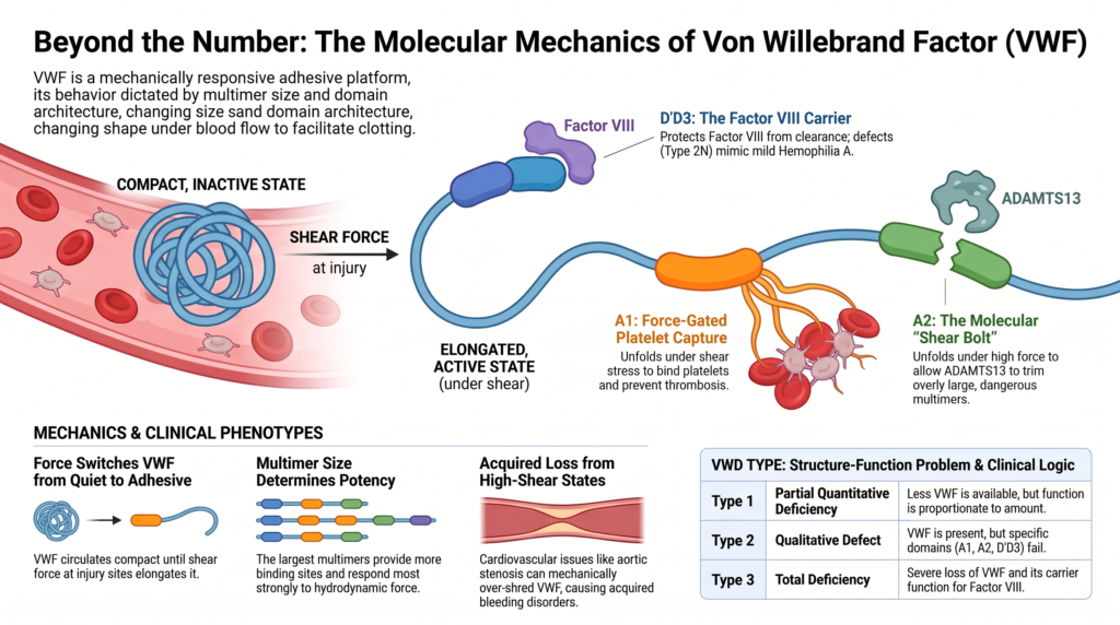
Von Willebrand factor is built to feel force.
That is the central structure-function lesson.
Von Willebrand factor (VWF) is not simply a soluble plasma protein. It is a multimeric, mechanically responsive adhesive platform whose multimer size, domain architecture, and force-dependent conformational change determine how it behaves in circulation and at sites of vascular injury. Glycosylation, storage, release, proteolysis, and clearance modify that behavior.1
This is why von Willebrand disease (VWD) is not just “low VWF.”
VWD can arise from defects in VWF synthesis, assembly, secretion, platelet binding, collagen binding, factor VIII binding, ADAMTS13 susceptibility, or clearance.
Clinical phenotypes emerge from which part of the VWF system fails.
Why this spoke matters
VWF structure explains why:
- VWF antigen and platelet-dependent VWF activity diverge
- high-molecular-weight multimers dominate function
- type 2 variants behave differently
- increased platelet binding can still cause bleeding
- type 2N can resemble mild hemophilia A
- desmopressin response depends on both release and survival
- high-shear cardiovascular states can cause acquired VWF dysfunction
- laboratory assays can mislead when they measure only one part of VWF biology
The clinician who understands VWF structure can interpret VWD as a mechanistic pattern, not simply a set of numbers.
VWF is assembled before it is released
VWF is encoded by the VWF gene on chromosome 12, which contains 52 exons and spans approximately 178 kb. It is synthesized in endothelial cells and megakaryocytes as pre-pro-VWF, a 2,813-amino acid precursor containing:2
- a 22-amino acid signal peptide
- a 741-amino acid propeptide, VWFpp
- a 2,050-amino acid mature VWF subunit
In the endoplasmic reticulum, VWF folds, forms disulfide bonds, receives N-linked glycans, and dimerizes through the C-terminal CK domain.
In the Golgi, additional glycan processing occurs, O-linked glycans are added, the propeptide is cleaved by furin, and VWF dimers multimerize through N-terminal D3-domain disulfide bonding.3
VWFpp is not merely discarded. It helps facilitate multimerization and regulated storage.4
Clinical implication:
A low VWF level may reflect reduced synthesis, impaired folding, defective multimer assembly, impaired secretion, increased clearance, or combinations of these mechanisms.
The antigen result does not tell you which step failed.
Domain architecture creates distinct failure modes
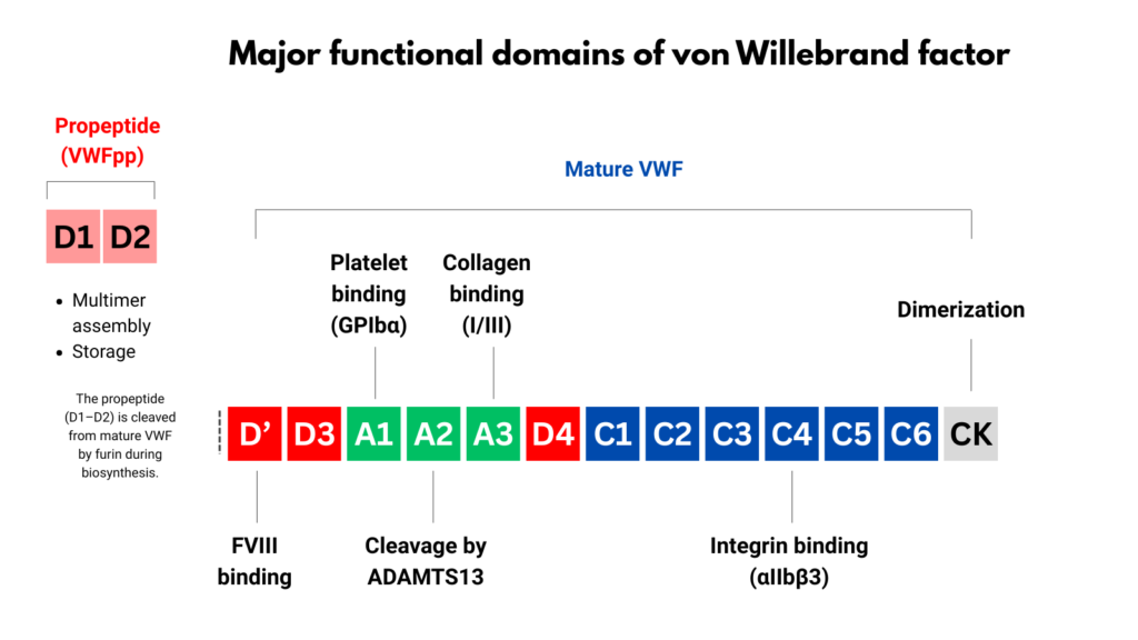
The mature VWF subunit is organized into repeated structural domains. The clinically useful domain map is:
D′-D3-A1-A2-A3-D4-C1-C2-C3-C4-C5-C6-CK
Modern structural annotation has refined some boundaries, especially in the D and C regions, but the classic map remains highly useful for clinical reasoning.5
Each domain represents a functional interface, and therefore a potential failure mode.
| Region | Major function | Clinical relevance |
|---|---|---|
| D′D3 | Factor VIII binding and stabilization | Type 2N VWD; low FVIII that can mimic mild hemophilia A |
| A1 | Platelet GPIbα binding; collagen IV/VI binding | Type 2B, type 2M, platelet-binding assays |
| A2 | ADAMTS13 cleavage site | Type 2A; VWF-ADAMTS13 axis; acquired shear-related VWF loss |
| A3 | Collagen I/III binding | Collagen-binding defects; selected type 2M phenotypes |
| C4 | RGD motif for platelet integrin αIIbβ3 | Platelet aggregation support |
| CK | C-terminal dimerization | Dimerization defects; multimer assembly failure |
Sources.6
Clinical implication:
Subtype classification reflects which structural interaction is disrupted, not merely how much VWF is present.
Multimer size is function
VWF circulates as a spectrum of multimers.
The largest multimers are disproportionately important for primary hemostasis because they provide multiple binding sites for collagen and platelets and are especially effective under shear.7
Large multimers matter for two reasons:
- they display more adhesive binding sites
- they respond more strongly to hydrodynamic force
Single-molecule and polymer-dynamics studies help explain why. In shear flow, force on VWF rises steeply with multimer length and is greatest toward the middle of the multimer. This gives larger multimers a mechanical advantage in unfolding, exposing binding sites, and capturing platelets.8
This advantage becomes most apparent under high shear, where smaller multimers may fail despite adequate antigen levels.
Clinical implication:
Two patients can have similar VWF antigen levels but different bleeding phenotypes if their multimer distributions differ.
That is why multimer analysis can change diagnosis.
Force switches VWF from quiet to adhesive
In intact vessels, VWF circulates in a compact, relatively inactive conformation. This helps prevent inappropriate platelet binding.
At sites of vascular injury, VWF binds exposed matrix and encounters hydrodynamic force. Under shear, VWF elongates, changes conformation, and exposes functional surfaces that allow platelet capture.9
Under force:
- VWF elongates
- platelet-binding function increases
- A1 becomes competent to bind platelet GPIbα
- A2 becomes susceptible to ADAMTS13 cleavage
- VWF supports platelet tethering and thrombus growth
This is the central paradox:
Shear activates VWF.
Shear also marks VWF for cleavage.
Shear is both activator and editor.
A1: force-gated platelet capture
The A1 domain mediates binding to platelet GPIbα.
This interaction is essential for platelet tethering under high shear, especially in the microcirculation and arterial circulation. But it must be tightly regulated. If VWF bound platelets freely in normal plasma, thrombosis would result.
A1 is therefore restrained in circulating VWF. Structural studies support the concept that flanking regions and domain organization help keep A1 from binding GPIbα inappropriately under resting conditions. Force-dependent conformational change releases this restraint and allows platelet binding.10
This structural logic produces two opposing type 2 phenotypes.
In type 2M VWD, VWF is present but platelet-dependent function is reduced. Multimers may be relatively preserved, but A1-mediated platelet binding is impaired.
In type 2B VWD, the problem is the opposite. Gain-of-function changes increase VWF affinity for platelet GPIbα. VWF binds platelets too readily, causing clearance of VWF-platelet complexes, loss of high-molecular-weight multimers, and sometimes thrombocytopenia.11
This is one of the most important structure-function lessons in VWD:
Both too little platelet binding and too much platelet binding can cause bleeding.
The mechanism differs, and so does management.
A2 and ADAMTS13: the VWF editing system
The A2 domain contains the ADAMTS13 cleavage site at Tyr1605-Met1606.
But the site is not freely available. In folded VWF, the cleavage site is buried. ADAMTS13 can cleave VWF efficiently only when force unfolds A2 enough to expose the scissile bond.12
This makes A2 a force-gated regulatory module.
A useful metaphor is that A2 is the shear bolt of VWF. A shear bolt is designed to give way above a defined force threshold to protect a larger system. Similarly, A2 unfolds under sufficient tensile force, exposing the ADAMTS13 cleavage site so that overly adhesive VWF multimers can be trimmed.13
The metaphor has limits. Unlike a mechanical bolt that breaks once, A2 unfolding and refolding are dynamic, force-dependent processes. But the concept is clinically useful: A2 is the structural element that allows VWF size and activity to be edited under flow.
ADAMTS13 is the enzyme that performs that editing. It circulates as an active protease, but it does not indiscriminately digest VWF because folded VWF protects the cleavage site. The key regulator is not simply whether ADAMTS13 is present. It is whether VWF has been mechanically presented in the right conformation.14
This paired system explains two extremes:
- too little VWF activity produces bleeding, as in VWD
- too little ADAMTS13 activity permits excessive ultra-large VWF activity, as in thrombotic thrombocytopenic purpura
The same regulatory axis helps explain opposite hemostatic consequences: loss of effective VWF function can produce bleeding, whereas severe ADAMTS13 deficiency permits pathologic ultra-large VWF activity and thrombosis.
Clinical implication:
Some type 2A variants reflect increased susceptibility of VWF to ADAMTS13-mediated cleavage, leading to loss of high-molecular-weight multimers and impaired platelet adhesion under shear.
Type 2A is therefore not one mechanism. It can reflect defective multimer assembly, defective secretion, increased ADAMTS13 susceptibility, or structural destabilization of multimers.15
Excess shear can create acquired VWF loss
The same force-dependent system can produce acquired VWF dysfunction.
In high-shear cardiovascular states, VWF may unfold excessively and become more susceptible to proteolysis. This can deplete high-molecular-weight multimers and produce an acquired type 2A-like pattern.
Aortic stenosis is the classic example. Severe aortic stenosis can be associated with mucocutaneous or gastrointestinal bleeding, loss of the largest VWF multimers, reduced collagen-binding activity, and correction after valve replacement when the shear abnormality is relieved.16
This is one mechanistic basis for Heyde syndrome, in which aortic stenosis, acquired VWF dysfunction, and gastrointestinal bleeding from angiodysplasia may coexist.
Similar principles apply in other high-shear settings, including some mechanical circulatory support devices.17
Clinical implication:
Loss of high-molecular-weight multimers can be inherited or acquired. The lab pattern may look similar, but the cause and treatment differ.
D′D3: constitutive factor VIII carriage
Not all VWF functions are force-gated.
VWF also serves as the carrier protein for factor VIII. The D′D3 region binds factor VIII with high affinity and protects it from premature clearance and proteolysis. Under normal conditions, most circulating factor VIII is bound to VWF.18
This explains two clinically important patterns.
In type 3 VWD, VWF is absent or nearly absent. Factor VIII may be markedly reduced because its carrier protein is missing. These patients may have both mucocutaneous bleeding and a more hemophilia-like bleeding pattern.
In type 2N VWD, VWF antigen may be normal or mildly reduced, but factor VIII binding is defective. The laboratory pattern may resemble mild hemophilia A because factor VIII is disproportionately low compared with VWF antigen.19
Clinical implication:
Type 2N is not “low VWF.” It is a carrier-function defect.
A patient with disproportionately low factor VIII should prompt consideration of type 2N VWD, particularly when the patient is female, the family pattern suggests autosomal inheritance, or VWF antigen is not proportionately reduced.
A3 and A1: collagen localization
VWF must bind platelets, but it must also localize to the damaged vessel wall.
Collagen binding provides this anchor.
The A3 domain binds primarily to fibrillar collagens I and III. The A1 domain contributes binding to collagens IV and VI.20
This creates a two-sided bridge:
- VWF binds exposed matrix
- VWF binds platelets in flowing blood
Unlike platelet GPIbα binding, which must be restrained until needed, collagen binding is more constitutively available. This allows VWF to localize rapidly to exposed subendothelial matrix.21
Clinical implication:
Some patients with bleeding, preserved antigen, preserved platelet-dependent activity, and normal multimers may still have impaired collagen binding. VWF collagen-binding assays may identify selected type 2M variants that are not specifically assessed by platelet-binding assays.22
Clearance is a structural phenotype
Clearance is not an afterthought. It is a major determinant of steady-state VWF levels and clinical phenotype.
Plasma VWF level reflects a balance among synthesis, secretion, proteolysis, and clearance. VWF clearance is influenced by glycosylation, conformation, macrophage uptake, lectin receptors, scavenger receptors, ABO blood group, and VWF sequence variants.23
VWFpp is released with mature VWF but has a much shorter half-life. The ratio of VWFpp to VWF antigen can help identify accelerated mature VWF clearance, although the assay is not universally available and interpretation requires context.24
ABO blood group is the best-known modifier. Blood group O is associated with lower VWF levels, in part through effects on VWF glycosylation and survival.25
Clinical implication:
A patient may have a good peak response to desmopressin but a short-lived response because released VWF is cleared rapidly. A desmopressin trial should assess both the height and duration of response.
Genotype does not always equal phenotype
The VWF gene is large, highly polymorphic, and complicated by a partial pseudogene on chromosome 22. Many variants occur in healthy individuals, and interpretation is affected by penetrance, modifier loci, ABO blood group, and the patient’s protein phenotype.26
Genetic testing is most useful when it clarifies a clinically important distinction, such as:
- type 2B VWD versus platelet-type VWD
- type 2N VWD versus mild hemophilia A
- type 2 subtype confirmation when phenotypic testing is equivocal
- type 3 VWD family planning
- congenital VWD versus acquired von Willebrand syndrome in selected cases
In type 1 VWD and low VWF phenotypes, genetic interpretation is more difficult. Many patients have no clearly pathogenic VWF variant, and non-VWF modifiers contribute to plasma VWF levels.28
Clinical implication:
Genetic testing supports classification, but it does not replace the protein phenotype, bleeding history, or clinical judgment.
Assays read structure imperfectly
VWF antigen measures how much VWF protein is present.
VWF activity assays estimate what VWF can do, most often platelet binding.
The relationship between antigen and activity is therefore a structure-function clue.
If VWF antigen and activity are proportionately reduced, the pattern supports a quantitative defect, such as type 1 VWD.
If activity is disproportionately reduced compared with antigen, the pattern suggests a qualitative defect, especially type 2 VWD.29
But assays have limitations.
Ristocetin-based assays do not reproduce physiologic shear. They use ristocetin as a surrogate to force VWF-GPIb interaction in vitro. Some A1 polymorphisms, especially D1472H, can reduce ristocetin-dependent activity without necessarily reducing in vivo VWF function or causing a bleeding phenotype.30
Newer GPIbM activity assays avoid ristocetin and may reduce some assay-related pitfalls, but static assays still do not fully reproduce VWF function under flow.31
Clinical implication:
A laboratory result is not the molecule. It is a constrained readout of one function under artificial conditions.
Subtype classification is structure-function shorthand
The modern VWD classification becomes more intuitive when viewed structurally.
| VWD type | Structure-function problem | Typical clinical logic |
| Type 1 | Partial quantitative deficiency | Less VWF is available, but function is usually proportionate to antigen |
| Type 1C | Accelerated clearance | VWF may be released normally but disappears too quickly |
| Type 3 | Near-complete or complete VWF deficiency | Severe loss of VWF, often with markedly reduced FVIII |
| Type 2A | Loss of high-molecular-weight multimers | Impaired platelet adhesion under shear |
| Type 2B | Increased A1-GPIbα binding | Platelet-VWF complex clearance, loss of HMW multimers, possible thrombocytopenia |
| Type 2M | Impaired platelet or collagen binding with relatively preserved multimers | VWF is present but does not bind normally |
| Type 2N | Impaired FVIII binding | Low FVIII may mimic mild hemophilia A |
Sources.32
This table is useful, but it should not be mistaken for biologic uniformity. Patients may sit at boundaries. Assays vary. VWF levels fluctuate with stress, inflammation, pregnancy, age, hormones, thyroid status, exercise, and blood group.33
Clinical implication:
Classification is a tool for reasoning, not a substitute for reasoning.
Structure guides treatment
Each therapy targets a different point in the VWF life cycle.
Desmopressin releases endogenous VWF from endothelial stores. It is most useful when the patient has stored, functional VWF that can be released and persists long enough to cover the hemostatic challenge.34
Desmopressin is most straightforward in many patients with type 1 VWD. It is ineffective in type 3 VWD, unreliable in many qualitative variants, and generally avoided or used only with expert guidance in type 2B because released abnormal VWF may worsen platelet binding and thrombocytopenia.35
A desmopressin trial should assess both peak level and duration of response. A brisk but short-lived response may suggest accelerated clearance and may not be adequate for prolonged bleeding-risk windows.
VWF concentrates replace missing or dysfunctional VWF. They are needed when endogenous VWF is absent, insufficient, unsafe to release, or qualitatively abnormal in ways that make desmopressin unreliable.
Factor VIII monitoring matters because VWF stabilizes FVIII. Some products contain both VWF and FVIII, while others are VWF-focused and require attention to FVIII kinetics.
Antifibrinolytics are especially useful for mucosal bleeding because mucosal surfaces have high fibrinolytic activity.36
The therapeutic logic follows the structural defect:
- release stored VWF when it is present and functional
- replace VWF when it is absent or dysfunctional
- support factor VIII when carrier function is inadequate
- stabilize mucosal clots when fibrinolysis is prominent
- avoid release strategies when released VWF may worsen platelet binding
Clinical synthesis
VWF structure explains why VWD is both simple and difficult.
It is simple because the core clinical problem is impaired hemostasis, usually mucocutaneous bleeding, sometimes combined with low factor VIII.
It is difficult because VWF is not a single-function protein. It is a force-responsive multimeric adhesive system.
At the bedside, structure-function reasoning asks four kinds of questions.
Quantity
- Is there enough VWF?
- Is the level stable over time?
Structure
- Are high-molecular-weight multimers present?
- Is VWF being cleaved or cleared too rapidly?
Function
- Does VWF bind platelets normally?
- Does it bind platelets too strongly?
- Does it bind collagen?
- Does it bind and protect factor VIII?
Interpretation
- Is the assay measuring biology or artifact?
- Does the bleeding phenotype fit the laboratory pattern?
The answer is rarely contained in one number.
Mechanism predicts phenotype.
Phenotype guides management.
Laboratory patterns must be interpreted structurally, not just numerically.
Final note
VWF is a molecule built for hemostasis in moving blood.
Its architecture allows it to circulate quietly, respond to force, bind injured vessel wall, recruit platelets, protect factor VIII, and be trimmed by ADAMTS13 before adhesion becomes excessive.
VWD occurs when one or more parts of that system fail.
The clinician who understands VWF structure can do more than classify disease. They can explain laboratory discordance, anticipate phenotype, recognize diagnostic pitfalls, and choose therapy with greater precision.
Evidence anchor: why structure-function reasoning matters in VWD
Summary derived from structural biology, mechanobiology, multimer studies, diagnostic reviews, mutation-spectrum studies, clearance literature, and management guidance. The evidence consistently shows that VWF function depends on domain-specific interactions, multimer size, force-dependent conformational change, ADAMTS13 proteolysis, and regulated clearance.
| Evidence stream | What it shows | Why it matters | Main limitation |
|---|---|---|---|
| Domain architecture | VWF domains map to distinct functions: D′D3 binds and stabilizes factor VIII; A1 mediates platelet GPIbα binding and contributes to collagen IV/VI binding; A2 contains the ADAMTS13 cleavage site; A3 binds collagen I/III; C4 contains the RGD motif for platelet integrin interaction; CK supports dimerization.37 | VWD subtypes are best understood as failures of specific structural functions, not simply as different degrees of “low VWF.” | Domain maps simplify a dynamic multimeric molecule and cannot fully capture conformation, shear, glycosylation, or cellular context. |
| Biosynthesis and storage | VWF is synthesized as pre-pro-VWF, dimerizes in the ER through the CK domain, multimerizes in the Golgi through N-terminal interactions, and is stored in Weibel-Palade bodies as ordered tubular structures.38 | Quantitative VWD can arise from defects in synthesis, folding, multimer assembly, storage, secretion, or clearance. Desmopressin response depends on stored and releasable endogenous VWF. | Most routine laboratory tests do not directly identify which biosynthetic step is abnormal. |
| Multimer biology | High-molecular-weight multimers are disproportionately effective in platelet adhesion and collagen interaction under shear.39 | Similar VWF antigen levels can have different clinical meaning if multimer distribution differs. Loss of high-molecular-weight multimers helps explain type 2A VWD and acquired high-shear VWF loss. | Multimer analysis is technically specialized, may not be rapidly available, and must be interpreted with other assays. |
| Mechanobiology and shear | Shear stress elongates VWF, promotes platelet-binding function, and exposes A2 to ADAMTS13 cleavage. Single-molecule studies support force-dependent A2 unfolding as a prerequisite for efficient ADAMTS13 cleavage.40 | VWF is built for hemostasis in moving blood. Shear both activates VWF and marks it for cleavage, explaining why VWF function cannot be fully inferred from static antigen measurements. | In vitro and single-molecule systems isolate mechanisms but do not fully reproduce the vascular microenvironment. |
| A1 platelet-binding phenotypes | Reduced A1-GPIbα interaction can produce type 2M-like platelet-binding defects, whereas increased A1-GPIbα binding causes type 2B VWD with VWF-platelet complex clearance, loss of high-molecular-weight multimers, and possible thrombocytopenia.41 | Both too little and too much platelet binding can cause bleeding. This distinction changes classification and influences desmopressin safety. | Platelet-binding assays are imperfect proxies for shear-dependent platelet capture in vivo. |
| A2-ADAMTS13 axis | ADAMTS13 regulates VWF multimer size by cleaving the A2 domain when the cleavage site is exposed by force. Too much cleavage can contribute to loss of high-molecular-weight multimers, while severe ADAMTS13 deficiency permits ultra-large VWF-mediated thrombosis.42 | VWD and TTP can be understood as opposite failures along the VWF-ADAMTS13 regulatory axis. A2 is the “shear bolt” that allows VWF size and activity to be edited under flow. | This axis explains many patterns but does not account for every modifier of bleeding or thrombosis risk. |
| High-shear acquired VWF loss | Severe aortic stenosis can cause acquired loss of the largest VWF multimers, reduced collagen-binding activity, and mucocutaneous or gastrointestinal bleeding, with improvement after valve replacement when shear is relieved.43 | A type 2A-like laboratory pattern can be acquired, not inherited. The same force-dependent biology explains inherited and acquired VWF dysfunction. | The presence of a high-shear lesion does not prove that all bleeding is VWF-mediated; clinical context remains essential. |
| Factor VIII carriage | The D′D3 region binds factor VIII and protects it from clearance. Type 2N VWD impairs factor VIII binding and can resemble mild hemophilia A.44 | Disproportionately low factor VIII should prompt consideration of type 2N VWD, especially when the inheritance pattern does not fit hemophilia A. | Factor VIII levels are influenced by multiple variables, so interpretation requires VWF antigen, activity, family pattern, and sometimes FVIII-binding or genetic testing. |
| Collagen-binding defects | VWF binds collagen I/III mainly through A3 and collagen IV/VI through A1. Some patients may have impaired collagen binding despite preserved antigen, platelet-dependent activity, and multimer distribution.45 | Collagen-binding assays can reveal selected type 2M mechanisms that platelet-binding assays may not specifically assess. | Collagen-binding testing varies by assay design and collagen type and is not uniformly available. |
| Clearance biology | Plasma VWF level reflects synthesis, secretion, proteolysis, and clearance. VWF clearance is influenced by glycosylation, macrophage uptake, lectin and scavenger receptors, ABO blood group, and VWF variants. The VWFpp/VWF:Ag ratio can help identify accelerated clearance.46 | A normal desmopressin peak may be misleading if VWF falls quickly. Duration of response matters for procedural planning. | VWFpp testing is not universally available, and clearance phenotypes may overlap with other quantitative defects. |
| Assay interpretation | VWF antigen, platelet-dependent activity, collagen binding, multimer analysis, factor VIII, VWFpp/VWF:Ag, and genetic testing are different windows into VWF biology. Ristocetin-based assays may be affected by assay-specific limitations such as A1 polymorphisms.47 | Laboratory results should be interpreted as partial structural readouts, not direct measurements of the whole molecule in vivo. | No single assay captures VWF function in moving blood, and results must be integrated with bleeding phenotype. |
Interpretive note: These evidence streams point in the same direction: VWF structure-function reasoning is not ornamental. It is the basis for interpreting antigen-activity discordance, multimer patterns, factor VIII levels, desmopressin response, acquired high-shear VWF loss, and subtype classification. The goal is not to memorize the domain map. The goal is to ask which structural function has failed.
Guideline synthesis: applying structure-function reasoning in VWD
Based on diagnostic reviews, laboratory guidance, genetic-testing discussions, and management guidance.
Use structure-function reasoning when interpreting:
- the lowest documented VWF antigen and platelet-dependent VWF activity levels
- whether antigen and activity are proportionate or discordant
- whether high-molecular-weight multimers are present
- factor VIII level relative to VWF antigen
- platelet count, especially when type 2B is possible
- collagen-binding results, if available
- VWFpp/VWF:Ag ratio or desmopressin survival curve, if clearance is suspected
- whether testing occurred during stress, inflammation, pregnancy, acute bleeding, or another VWF-raising state
- whether the bleeding history fits the laboratory pattern
- whether the patient has faced meaningful hemostatic challenges
- whether a planned procedure requires a treatment plan even if the label remains uncertain
Avoid common interpretive errors
- treating VWF antigen as the whole diagnosis
- assuming normal antigen excludes qualitative VWD
- assuming low platelet-dependent activity always means type 1 VWD
- missing type 2N VWD in a patient labeled as mild hemophilia A
- missing type 2B VWD when thrombocytopenia and loss of high-molecular-weight multimers are present
- judging desmopressin response only by the peak value
- treating multimer analysis as optional trivia when type 2A or 2B is suspected
- interpreting ristocetin-based activity as if it perfectly reproduced shear-dependent VWF function
- assuming acquired VWF loss cannot mimic inherited type 2A patterns
- letting subtype terminology replace mechanism-based reasoning
Useful language
Instead of:
“Your VWF antigen is not that low.”
Try:
“Your VWF amount is only one part of the picture. We also need to know how well the VWF works, whether the largest multimers are present, and whether the bleeding history fits.”
Instead of:
“The activity is low, so this is type 1.”
Try:
“When activity is lower than antigen, we should think about a qualitative VWF problem, not just a quantitative deficiency.”
Instead of:
“The desmopressin trial worked.”
Try:
“The peak response was good. Now we need to know whether the response lasted long enough for the bleeding-risk situation.”
Instead of:
“This looks like mild hemophilia A.”
Try:
“Because factor VIII is disproportionately low, we should consider whether VWF is failing to bind and protect factor VIII, as in type 2N VWD.”
Instead of:
“The multimer result is just a specialty test.”
Try:
“The multimer pattern tells us whether the VWF present has the size distribution needed for platelet adhesion under shear.”
Instead of:
“The aortic stenosis is unrelated to the bleeding workup.”
Try:
“High shear can cause acquired loss of large VWF multimers, so the valve lesion may be part of the hemostatic explanation.”
Practical takeaway
Structure-function reasoning makes VWD safer to interpret.
The question is not only “How much VWF is there?” It is:
- What size is it?
- What does it bind?
- How does it behave under force?
- Is it being cleaved or cleared too quickly?
- Does the laboratory pattern explain the bleeding phenotype?
- Does the mechanism predict the treatment plan?
The best VWD care interprets VWF as a system, not a number.
Reflect & Apply Case
A patient has:
- normal VWF antigen
- normal multimer distribution
- markedly reduced platelet-binding activity
Which structural interaction is most likely impaired?
This pattern points toward impaired interaction between the A1 domain and platelet GPIbα. Because multimer distribution is preserved, the mechanism resembles type 2M VWD more than classic type 2A VWD.
The lesson is not simply that the activity is low.
The lesson is that the VWF protein is present, but its platelet-binding function is impaired.
That is structure-function reasoning.
Test your thinking
A short quiz on VWF structure and function.